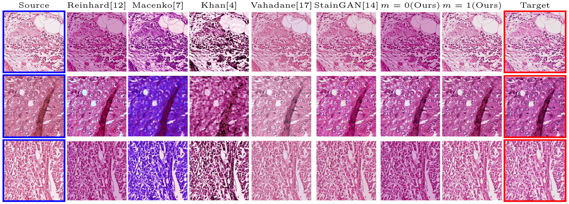
Fig.3.

Multimarginal Wasserstein Barycenter comparison with state of the art methods on MITOS-ATYPIA’14 challenge dataset. The blue bordered image is the input image and the two red bordered images are the references (with the one above being the intermediate reference). m is the number of intermediate references (in traditional Wasserstein barycenter m = 0; there is no intermediate reference).
