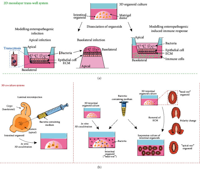
Figure 2.

In vitro modeling of enteropathogenic infection. (a) 2D intestinal coculture models: bacteria are seeded onto the apical or basolateral surface of the intestinal epithelial monolayer (adapted from: Ranganathan et al., 2019 [38], Koestler et al., 2019 [39]). Optionally, immune cells are added to the basolateral compartment of infected intestinal epithelium (adapted from: Noel et al., 2017 [31], Karve et al., 2017 [49]). (b) 3D intestinal coculture models: bacteria are either introduced into intestinal organoids via luminal microinjection (adapted from: Karve et al., 2017 [49]) or added to the culture medium of “basal-out” or “apical-out” intestinal organoids (adapted from: Co et al., 2019 [59]).
