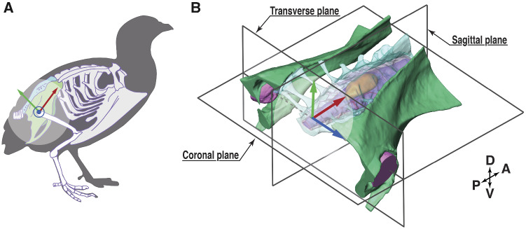
Fig. 1.
The anatomy of the synsacrum of the quail. (A) Schematic skeletal outline of a quail synsacrum, emphasized in green and turquoise. The schematic was modified from Modesto (2009), CC license. (B) A 3D view of the synsacrum, also showing a local coordinate system (x-red, y-green, and z-blue, used in “Digital dissection, uCT scanning” section), and three planes of reference. The coordinate origin is located at the center between both femur head sockets, with the coronal plane in-parallel to the orientation of the denticulate ligament network (Fig. 2). Abbreviations are explained in Appendix Table A3.

