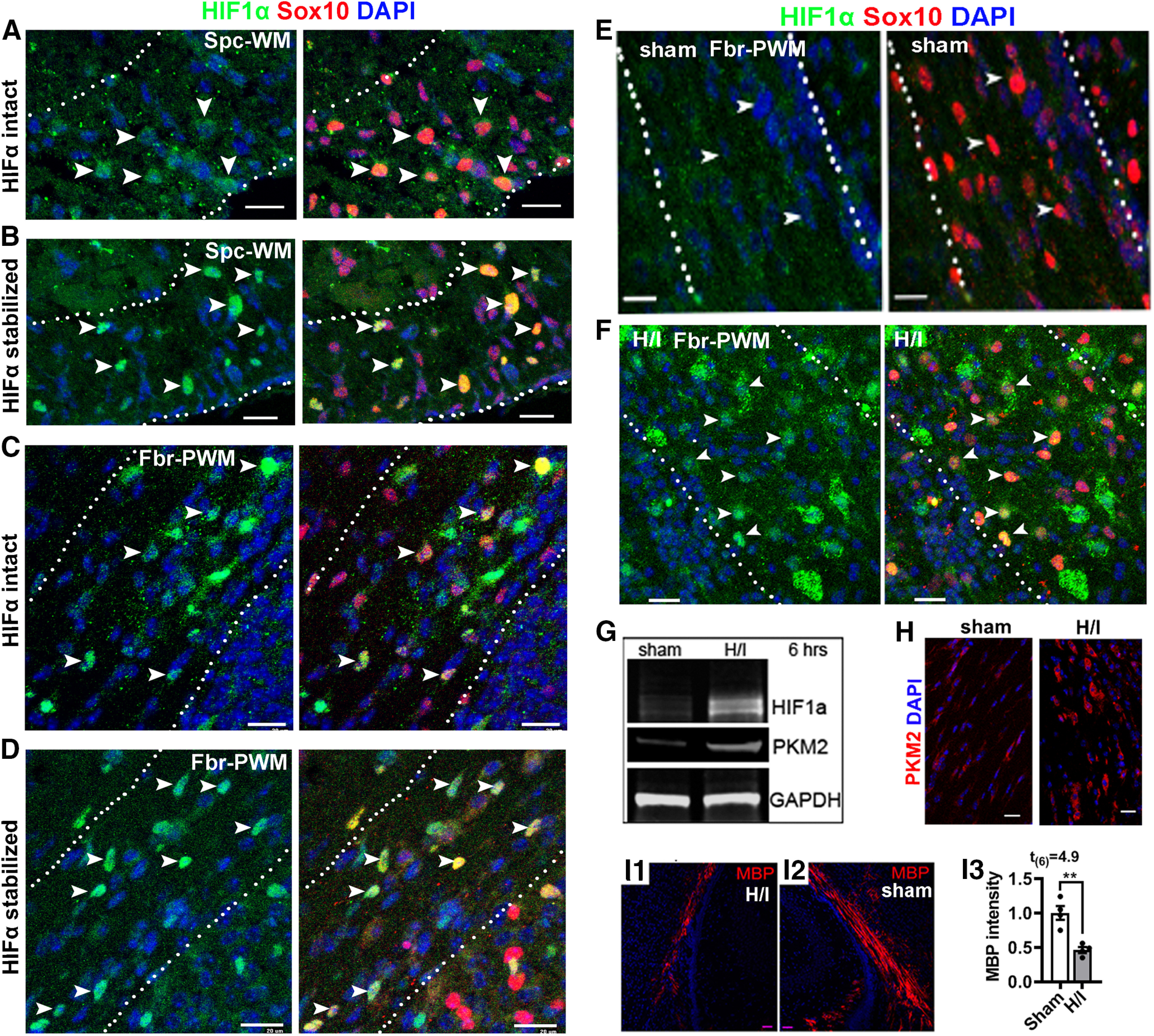
Figure 1.

HIFα stabilization in the early postnatal murine CNS. A, B, IHC of HIF1α and oligodendroglial lineage marker Sox10 in the spinal cord ventral white matter (Spc-WM, marked by dotted lines) of HIFα intact mice (A) and HIFα stabilized (Sox10-Cre:Vhlfl/fl) mice (B) at P5. C, D, IHC of HIF1α and Sox10 in the forebrain periventricular white matter (Fbr-PWM; marked by dotted lines) of HIFα intact mice (C) and HIFα stabilized (Sox10-Cre:Vhlfl/fl) mice (D) at P5. E, F, IHC of HIF1α and Sox10 in the Fbr-PWM of P10 mice that had been subjected to sham operation (E) and H/I injury on P6 (i.e., 4 d post-H/I or sham; F; for details, see Materials and Methods). Arrowheads in A–F point to HIF1α+Sox10+ cells. G, Western blot of microdissected subcortical white matter for HIF1α and canonical HIFα target PKM2 at 6 h post-H/I or sham. GAPDH, Internal loading control. H, PKM2 IHC in the Fbr-PWM at P10, 4 d post-H/I or sham. I1–I3, IHC and quantification of MBP staining in the Fbr-PWM at P10, 4 d post-H/I or sham. MBP intensity was normalized to that of the contralateral brain hemispheres to the occluded artery of the same mouse. Scale bars, 20 µm.
