Figure 10.

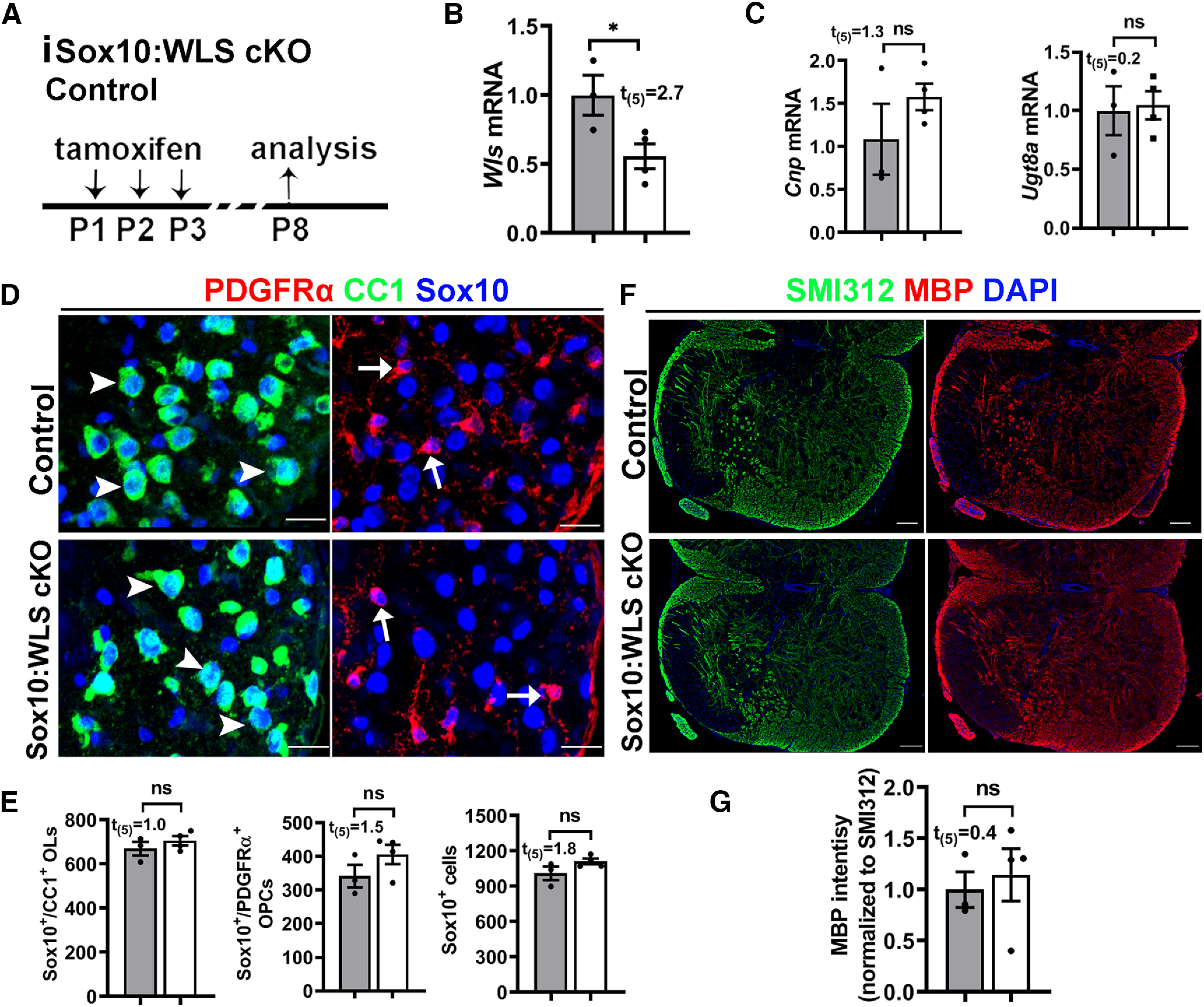
Disrupting WLS in Sox10-expressing oligodendroglial lineage cells does not perturb normal OPC differentiation and myelination. A, Experimental design for B–G. Tamoxifen-inducible Sox10-CreERT2:Wlsfl/fl (iSox10:WLS cKO, n = 4); littermate non-Cre control Wlsfl/fl mice (n = 3). B, C, qRT-PCR quantification of Wls (B) and differentiated OL-enriched gene Cnp and Ugt8a (C) in the spinal cord. D, E, Representative confocal images (D) and quantification (E) of Sox10+/CC1+ differentiated OLs (D, arrowheads), Sox10+/PDGFRα+ OPCs (D, arrows), and Sox10+ oligodendroglial lineage cells in the spinal cord. F, G, Representative confocal images (B) and quantification (C) of myelination by MBP staining of the spinal cord. SMI312 signal was used as an internal control of MBP quantification. Scale bars: D, 20 µm; E, 50 µm.
