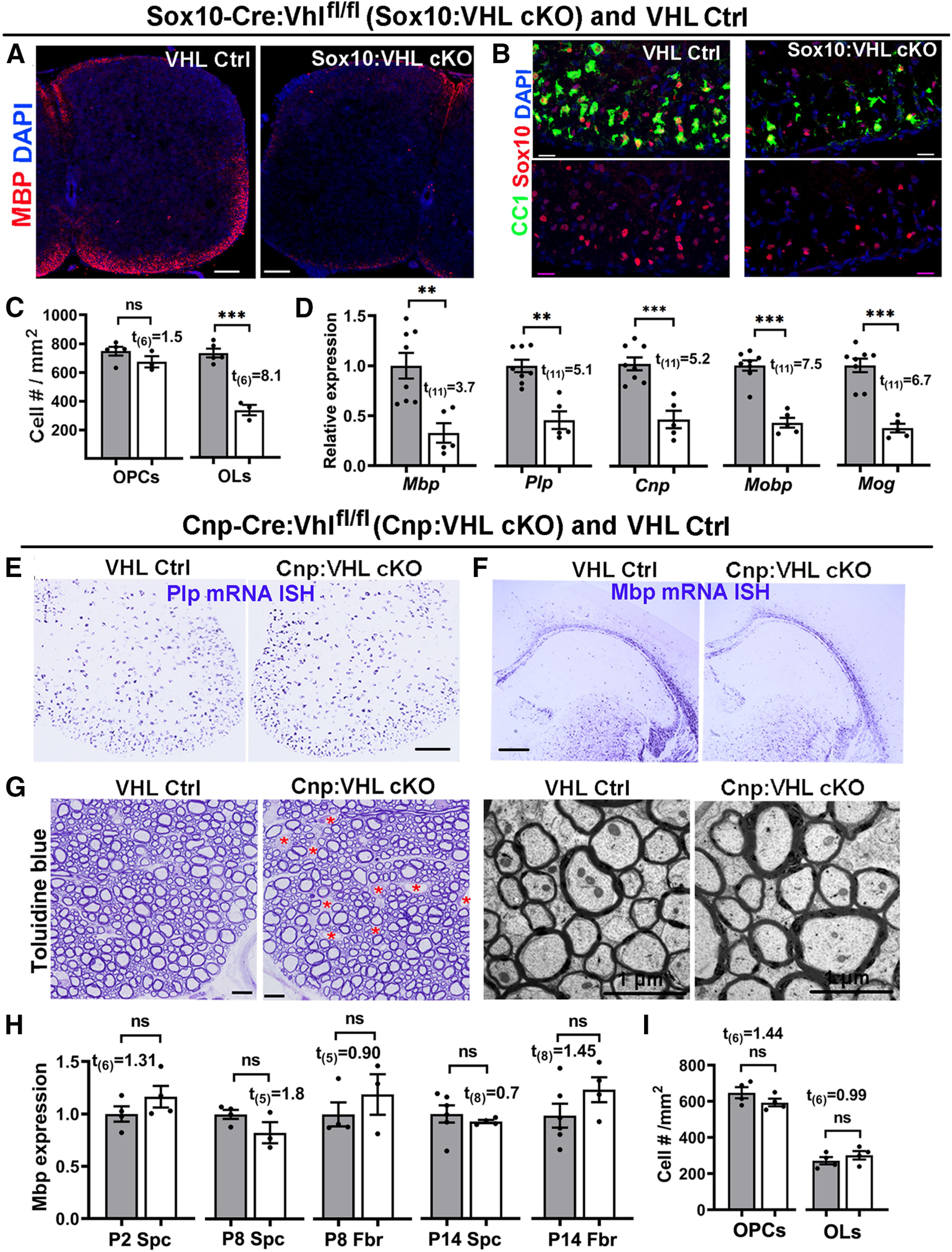
Figure 2.

HIFα activation impairs developmental myelination by inhibiting OPC differentiation but not subsequent OL maturation. A, Myelin staining of MBP in the spinal cord of Sox10-Cre:Vhlfl/fl (Sox10:VHL cKO) and littermate Ctrl (Vhlfl/+ or Vhlfl/fl) pups at P5. Most Sox10:VHL cKO pups died by the first postnatal week. B, Representative confocal images of Sox10 (red) and differentiated OL marker CC1 (green) in P5 spinal cord (Ctrl, n = 5; cKO, n = 3). Blue, DAPI nuclear counterstaining. C, Quantification of Sox10+CC1+ OLs and Sox10+PDGFRa+ OPCs (right) in P5 spinal cord (n = 5 Ctrl, n = 3 cKO). D, qRT-PCR assay for myelin-specific genes in the spinal cord at P5. Plp, Proteolipid protein; Cnp, 2′,3′-cyclic-nucleotide 3′-phosphodiesterase; Mobp, myelin oligodendrocyte basic protein; Mog, myelin oligodendrocyte glycoprotein. E, F, mRNA in situ hybridization (ISH) of Plp and Mbp in the spinal cord (E) and forebrain (F) of Cnp-Cre:Vhlfl/fl (Cnp:VHL cKO) and littermate control (Cnp-Cre:Vhlfl/+, Vhlfl/+, or Vhlfl/fl) mice at P14. G, Myelination in the adult spinal cord indicated by toluidine blue staining of semithin sections (left) and TEM of ultrathin section (right). Red asterisks in G denotes blood vessels, the density of which is elevated in Cnp:VHL cKO mice (Guo et al., 2015). H, Mbp mRNA levels quantified by qRT-PCR in Cnp:VHL cKO and Ctrl mice at different time points. Spc, Spinal cord; Fbr, forebrain. I, densities of Sox10+CC1+ OLs and Sox10+PDGFRα+ OPCs in P8 spinal cord of Cnp:VHL cKO and Ctrl mice. Scale bars: A, 100 µm; B, 20 µm; E, F, 200 µm; G, left, 10 µm; G, right, 1 µm. Data are presented as the mean ± SEM; gray bars are from Ctrl, white bars from cKO; two-tailed Student's t test, *p < 0.05; **p < 0.01; ***p < 0.001; ns, not significant. The above data presentation and statistics are applied to all figures unless otherwise indicated).
