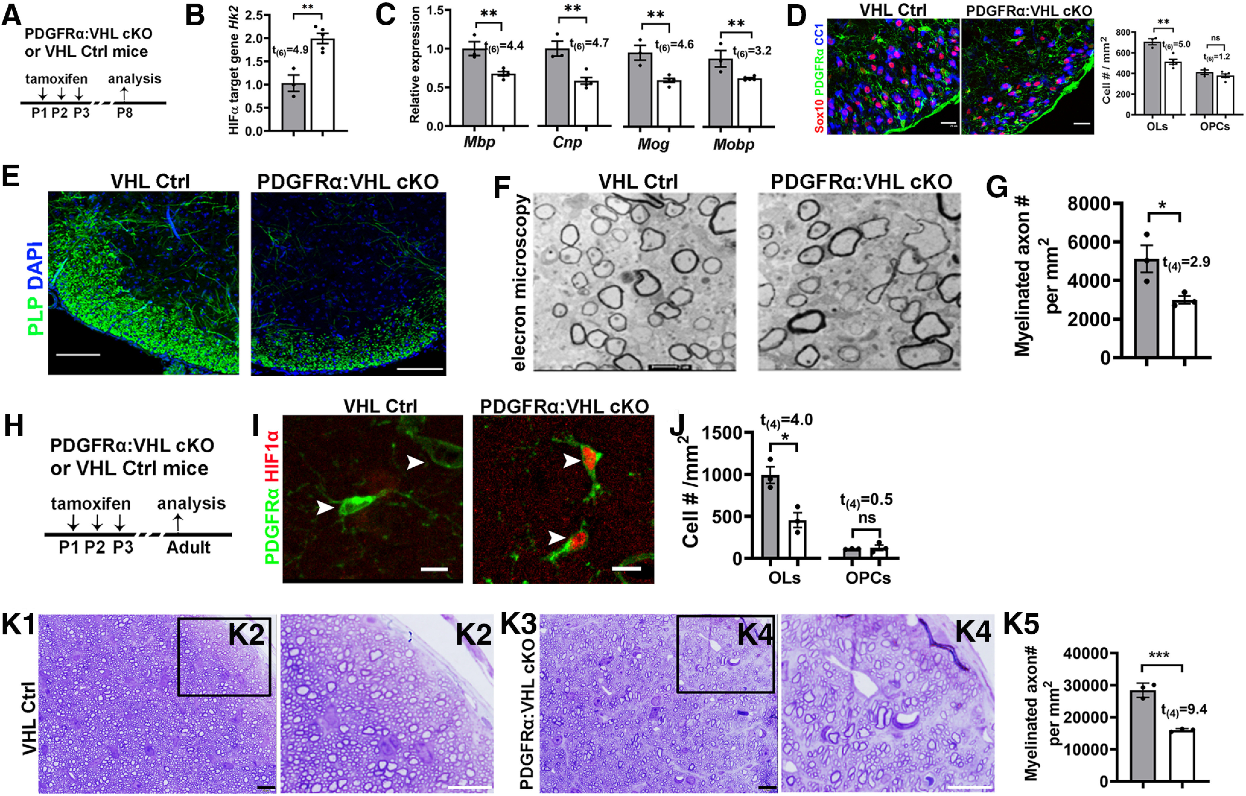
Figure 3.

Inducible HIFα stabilization in postnatal OPCs inhibits OPC differentiation. A, experimental design for B–F. PDGFRα:VHL cKO (Pdgfrα-CreERT2:Vhlfl/fl) and littermate control (Vhlfl/fl) mice were treated with tamoxifen intraperitoneally at P1, P2, and P3, and analyzed at P8. B, qRT-PCR quantification of HIFα target gene hexokinase 2 Hk2 in the spinal cord. C, Expression of major myelin proteins in the spinal cord quantified by qRT-PCR. D, Representative confocal images and the density of Sox10+/CC1+ OLs and Sox10+/PDGFRα+ OPCs. E, IHC of PLP showing myelination in the spinal cord ventral white matter. F, G, Representative TEM images (F) and the density of myelinated axons (G) in the spinal cord ventral white matter. H, Experimental design for I–K. PDGFRα:VHL cKO and littermate control mice were treated with tamoxifen intraperitoneally at P1, P2, and P3, and analyzed at P60–P70. I, Representative confocal images showing HIF1α stabilization in PDGFRα+ OPCs (arrowheads) of PDGFRα:VHL cKO mice. J, Densities of CC1+ mature OLs and PDGFRα+ OPCs in the corpus callosum. K1–K4, Toluidine blue staining for myelin on semithin (500 nm) resin sections showing hypomyelination in the optic nerve of adult PDGFRα:VHL cKO mice. Boxed areas in K1 and K3 were shown at higher-magnification images in K2 and K4, respectively. K5, Density of myelinated axons in the adult optic nerves. Scale bars: D, 25 µm; E, 100 µm; F, 0.5 µm; I–K5, 10 µm.
