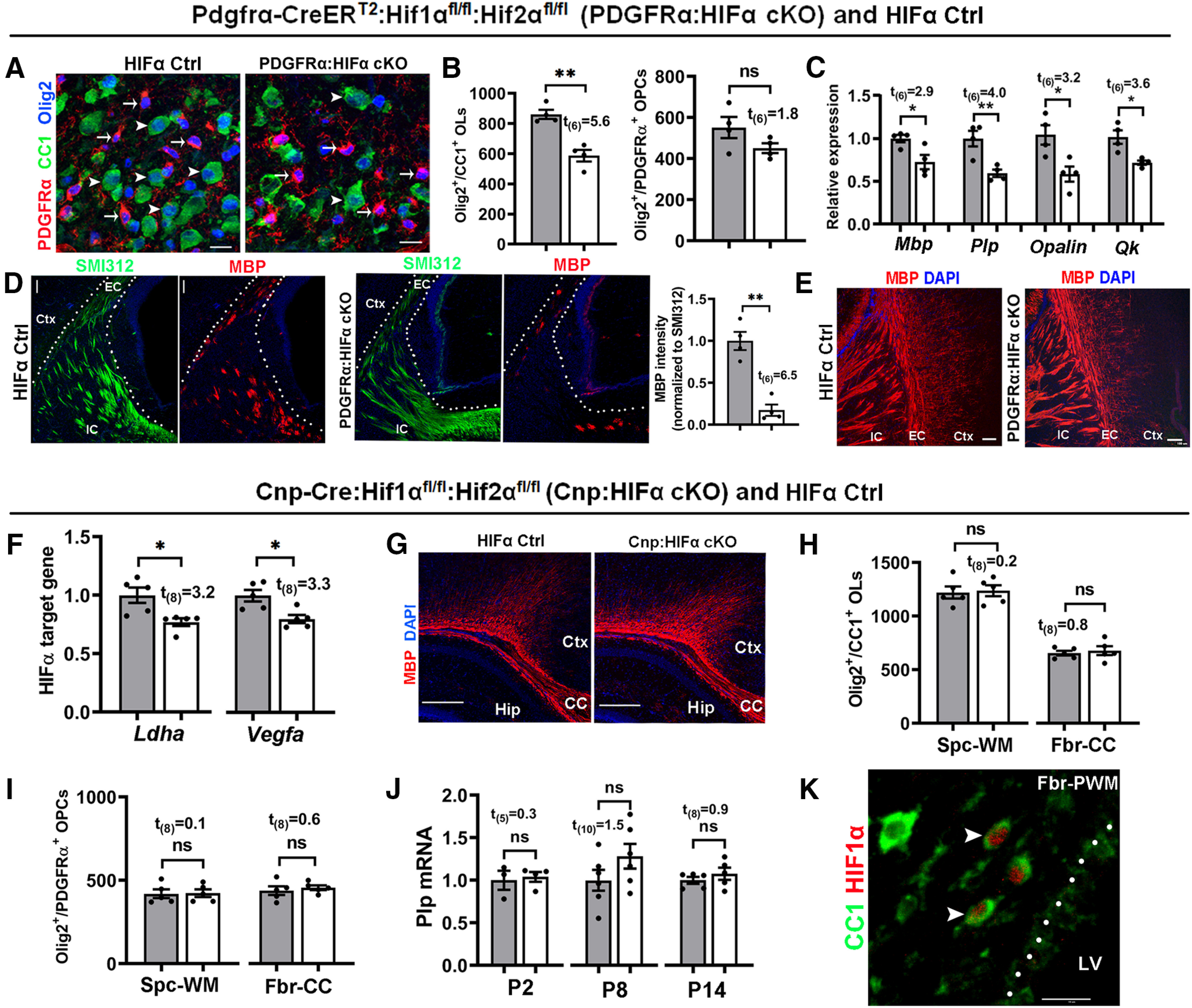
Figure 4.

HIFα inactivation transiently delays developmental myelination by controlling OPC differentiation but not subsequent OL maturation. A–E, PDGFRα:HIFα cKO and littermate control (Hif1αfl/fl:Hif2αfl/fl) mice were treated with tamoxifen at P1, P2, and P3, and analyzed at P8 (n = 4 each group) and P14 (n = 3 each group). A, B, IHC (A) and quantification (B) of Olig2+CC1+OLs (arrowheads) and Olig2+PDGFRα+ OPCs (arrows) in the spinal cord. C, Relative expression of myelin protein genes of Mbp and Plp and mature OL-specific gene of Opalin and quake [Qk (called CC1)] in the forebrain measured by qRT-PCR. D, IHC of myelin marker MBP and pan-axonal marker SMI312 in forebrain periventricular white matter (left, marked by dotted area) and quantification (right) at P8. EC, External capsule; IC, internal capsule; Ctx, cortex. E, IHC of MBP in forebrain periventricular white matter at P14 (tamoxifen at P1, P2, and P3). F–J, Cnp:HIFα cKO and littermate control (Hif1αfl/fl:Hif2αfl/fl) mice were analyzed at P2, P8, and P14. F, qRT-PCR quantification of HIFα target genes Ldha and Vegfa in P8 spinal cord. G, Myelin staining by MBP in the forebrain. CC, Corpus callosum; Hip, hippocampus. H, I, Densities (#/mm2) of Olig2+CC1+ OLs (H) and Olig2+PDGFRα+ OPCs (I) at P14. Spc-WM, Spinal cord white matter; Fbr-CC, forebrain corpus callosum. J, qRT-PCR assay of exon3b-containing Plp mRNA, which is specific to mature OLs, in the spinal cord at different time points. K, IHC of CC1 and HIF1α in P8 forebrain periventricular white matter (Fbr-PWM). Arrowheads point to double-positive cells. LV, Lateral ventricle. Scale bars: A, K, 10 μm; D, 20 μm; E, 100 μm; G, 250 μm.
