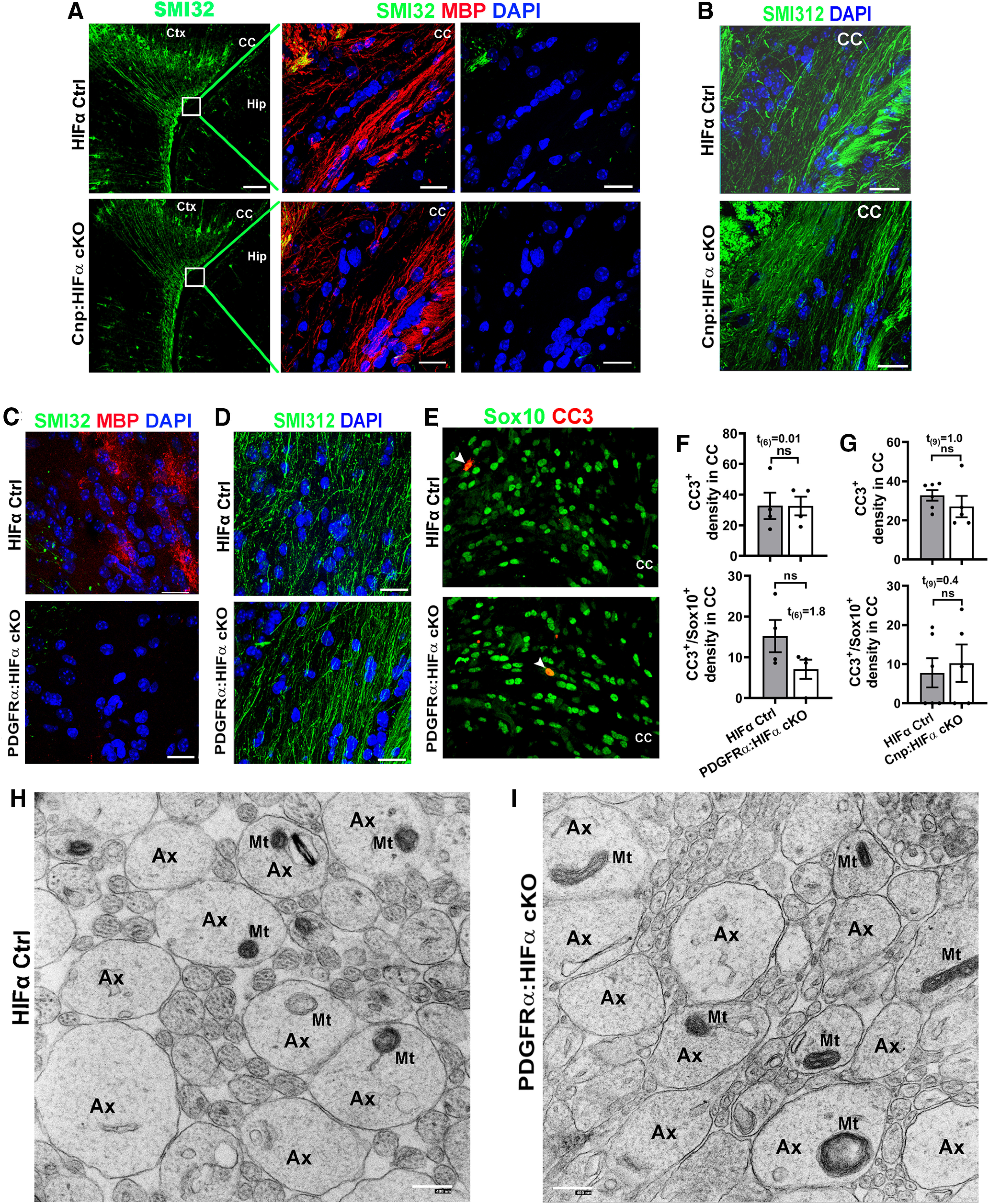
Figure 5.

Oligodendroglial HIFα is dispensable for axonal integrity or cell death. A, Immunostaining images of P14 forebrain using SMI32, a monoclonal antibody recognizing the nonphosphorylated neurofilament proteins of heavy chain (NFH), which has been reported labeling injured axons and a subset of pyramidal neuron bodies and dendrites. Boxed areas show the corpus callosum (CC) in higher-magnification images of SMI32 and MBP in the right panels. Ctx, Cortex; Hip, hippocampus. Note the absence of SMI32-positive signals in the CC of both Cnp:HIFα cKO and Ctrl mice. B, Immunostaining images of P14 CC using SMI312, a monoclonal antibody recognizing both phosphorylated and nonphosphorylated NFH (a pan-axonal marker). C, D, IHC of SMI32/MBP (C) and SMI312 (D) in the CC of P8 non-Cre Ctrl and PDGFRα:HIFα cKO and littermate control (Hif1αfl/fl:Hif2αfl/fl) mice (tamoxifen injection at P1, P2, and P3). E, F, Representative confocal images and quantification of cells positive for cleaved CC3 and/or pan-oligodendroglial lineage marker Sox10 in P8 CC of PDGFRα:HIFα cKO and control mice. Arrowheads point to CC3+/Sox10+ cells. G, Quantification of CC3+ and CC3+/Sox10+ cells in P14 CC of Cnp:HIFα cKO and control mice. H, I, Representative TEM images showing the cross-sections of axons in the corpus callosum of HIFα Ctrl (I) and cKO (H) mice at P8 (tamoxifen injections at P1, P2, and P3) when myelination is barely detectable at the TEM level. Note that the morphology of axons (Ax) and axonal mitochondria (Mt) were indistinguishable between HIFα Ctrl and cKO mice. Scale bars: A, low magnification, 100 µm; A, high magnification, 20 µm; B–E, 20 µm; H, I, 400 nm.
