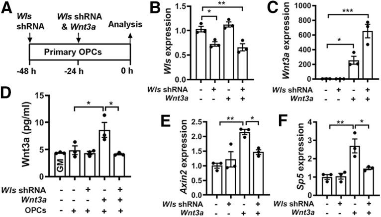
Figure 7.

WLS deficiency inhibits Wnt3a secretion and blocks autocrine Wnt/β-catenin signaling activity in OPCs. A, Experimental design of cell transfection of Wls shRNA and Wnt3a-expressing plasmids to primary brain OPC maintained in the growth medium. B, C, qRT-PCR assay of Wls (B) and Wnt3a (C) expression in transfected primary OPCs. One-way ANOVA followed by Tukey's multiple-comparisons test: Wls, F(3,8) = 15.39, p = 0.001; Wnt3a, F(3,8) = 32.81, p < 0.0001. D, ELISA quantification of Wnt3a in the growth medium in the absence or presence of OPCs transfected by Wls shRNA and/or Wnt3a. One-way ANOVA followed by Tukey's multiple-comparisons test, ***p < 0.001; ns, not significant. F(4,10) = 6.70, p < 0.0069. Note that Wnt3a concentration in the GM in the presence of primary OPCs is statistically indistinguishable from that in the GM alone, suggesting that intact primary OPCs do not secrete Wnt3a. E, F, qRT-PCR quantification of Wnt target genes Axin2 and Sp5 in OPCs transfected with Wls shRNA and/or Wnt3a. One-way ANOVA followed by Tukey's multiple-comparisons test: Axin2, F(3,8) = 11.06, p = 0.0032; Sp5, F(3,8) = 13.25, p = 0.0018.
