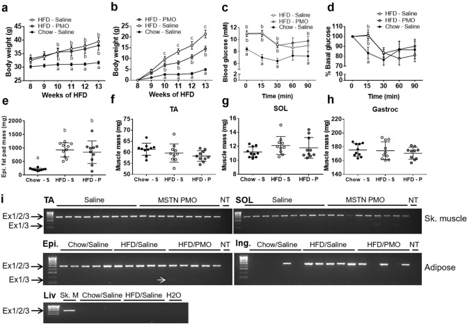
Figure 5.
Intravenous administration of unmodified PMOs does not induce sufficient exon skipping to increase muscle mass or decrease fat mass in high-fat diet-fed mice. (a,b) Body mass (a) and percentage increase in body mass (b) measured weekly after the start of i/v PMO or saline administration (start: week 8). (c,d) Absolute (c) and relative (d) blood glucose concentrations during the IPITT. E–H: Epididymal fat pad (e), TA muscle (f), SOL muscle (g) and Gastrocnemius (h) muscle masses in saline-treated chow-fed (Chow-S), saline-treated HFD-fed mice (HFD-S) and PMO-treated HFD-fed mice (HFD-P). (i) RT-PCR/agarose gel analysis of myostatin exon skipping in muscle, adipose and liver tissue. Epi: epididymal fat pad. Ing: inguinal fat pad. Liv: Liver; NT: non-template control. Ex1/2/3: Unskipped myostatin transcript; Ex1/3: Skipped myostatin transcript. Small white arrow indicates PCR product from skipped myostatin mRNA. Data are shown as mean ± S.E.M (a–d), with different letters indicating significant differences between the groups at each time point (p < 0.05), or as individual data points and means ± S.D. (E–H). N = 10 per group.

