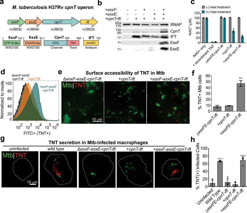
Fig. 1. EsxE-EsxF are required for TNT surface accessibility and secretion by M. tuberculosis.
a The cpnT operon of Mtb and the domain organization of the encoded proteins. TNT tuberculosis necrotizing toxin, IFT immunity factor to TNT, NTD N-terminal domain. b CpnT protein levels are dependent on EsxE and EsxF. Immunoblot of Mtb whole-cell lysates detected by antibodies specific for the indicated proteins. CpnT was detected using an anti-TNT antibody for the C-terminal domain. RNA polymerase (RNAP) was used as a loading control. Representative of two experiments. c The NAD+ glycohydrolase activity of Mtb is dependent on EsxE and EsxF. The NAD+ glycohydrolase activity of whole-cell lysates of Mtb strains was determined without or with heat treatment at 65 °C to release the antitoxin from TNT. The residual NAD+ concentration was measured by conversion of NAD+ to a fluorescent intermediate after NaOH treatment. NAD+ with only buffer and with added recombinant TNT were used as negative and positive controls, respectively. Representative experiment shown from two separate trials. d, e Surface accessibility of TNT of the indicated Mtb strains by flow cytometry using an α-TNT antibody and FITC-conjugated secondary antibody (d) and by fluorescence microscopy of Mtb strains stained with DMN-trehalose (green), and probed with α-TNT and Alexafluor-594-conjugated secondary antibody (e). f Quantification of e. Percentage of TNT-positive cells out of total bacteria from at least five fields of view. N ≥ 1000 bacterial cells total over two independent experiments. Shown is mean and standard deviation from two experiments. The statistical analysis was performed using one-way ANOVA analysis with Dunnet’s multiple comparison test using the ΔesxFE-cpnT-ift strain as the negative control. P = 0.0018. g Detection of TNT in the cytosol of Mtb-infected macrophages is dependent on EsxE and EsxF. Shown are THP-1 macrophages 48 h after infection with Mtb. THP-1 cells were treated with digitonin to only permeabilize the plasma membrane and were probed with anti-TNT antibody (Alexafluor-594, red). h Quantification of g. Infected macrophages were scored as TNT+ or TNT− based on the presence or absence of bright red punctae and quantified as % TNT-positive cells. At least 100 cells were analyzed of at least four independent experiments. Data shown is the mean ± SD for four biological replicates. Statistical analysis was performed using one-way ANOVA with Dunnet’s multiple comparison test compared to the uninfected sample as the negative control. P < 0.0001. Source data are provided in the Source data file.

