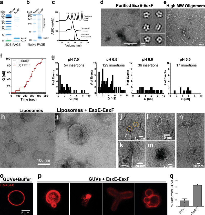
Fig. 4. Pore formation by the EsxE-EsxF complex and interaction with membranes.
a Coomassie-stained SDS–polyacrylamide gel and native polyacrylamide gel (b) of the purified EsxE-EsxF complex after anion exchange chromatography (representative of more than three purifications). The individual purification steps are shown in Fig. S4. c Gel filtration of EsxE-EsxF under native and denaturing conditions indicating oligomerization. d Electron microscopy of the EsxE-EsxF complex stained with 1% uranyl formate. Shown are reference-free 2D class averages from 19,919 particles. The full class averages are found in Fig. S7. Data are from a single EM session. Representative of at least three separate experiments. e Electron micrograph of the high molecular weight oligomers of EsxE-EsxF (filaments) negatively stained with 1% uranyl acetate. Representative of at least two separate experiments. f Current trace of EsxE-EsxF in diphytanoyl phosphatidylcholine bilayers in 25 mM NaPO4 (pH 6.5), 1 M KCl. A total of 1 µg protein was added to both cis and trans compartments, and 10 mV voltage was applied. Results are representative of three protein preparations over ten membranes. g Total channel insertion events (membrane pore formation) shown as the number of events as a factor of the conductance values observed at different pH values. Results are representative of at least ten membranes of three independent experiments. The complete profiles with the histograms, channel openings and closings for each condition are shown in Fig. S6. h, i Negative stain electron micrographs of dimyristoyl-phosphatidylcholine liposomes with (i) or without (h) EsxE-EsxF for 30 m at room temperature in 25 mM sodium phosphate (pH 6.5), 150 mM NaCl stained with 1% uranyl formate. The final EsxE-EsxF protein concentration was 50 nM. Representatives of two separate experiments shown. j–m Selected events indicating EsxE-EsxF membrane binding or interaction with liposomes. Inset in k. Enlarged membrane pore of EsxE-EsxF. m Aggregates pores. n EsxE-EsxF filaments binding to liposomes. o Interaction of EsxE-EsxF with membranes. Giant unilamellar vesicles (GUVs) were treated with the dye FM464X to label the lipid membranes (o), and were then incubated with 40 µg EsxE-EsxF (p) in a buffer containing 25 mM sodium phosphate (pH 6.5) and 150 mM NaCl. q Quantification of the number of deformed GUVs in the presence of EsxE-EsxF. Non-spherical objects which retained the FM464X dye were defined as deformed. The total number of GUVs from two independent experiments was 78 GUVs in buffer and 73 “GUVs” after incubation with EsxE-EsxF. The values are shown as the mean ± standard deviation. Source data are provided in the Source data file.

