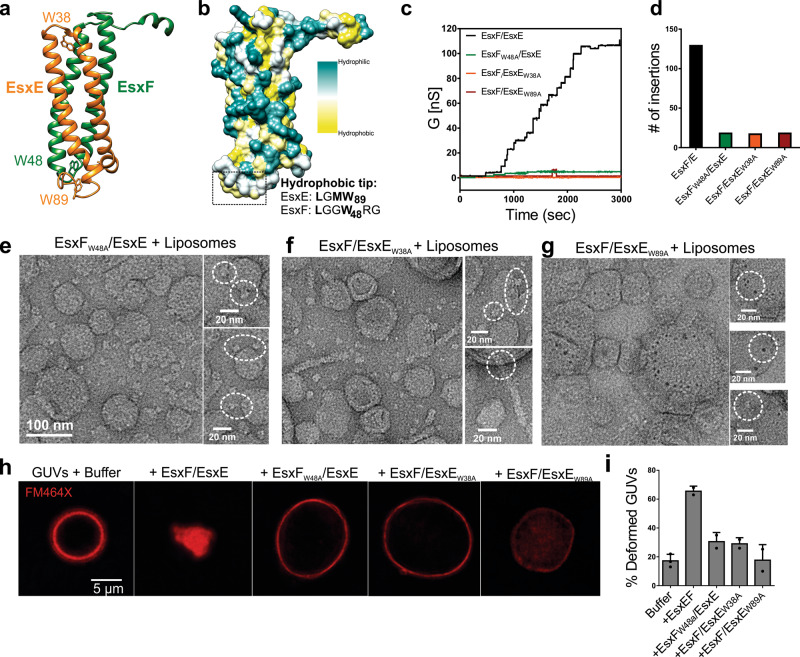
Fig. 5. The WXG motif is required for pore-forming activity.
a Homology model of EsxE-EsxF indicating the location of the WXG motifs and the EsxE C-terminal GXW motif. The respective tryptophans are highlighted. b Hydrophobicity plot of the EsxE-EsxF complex. Some of the residues forming the hydrophobic tip are highlighted. c Current trace of wt EsxE-EsxF and the WXG variants in 25 mM sodium phosphate (pH 6.5), 1 M KCl in diphytanoyl phosphatidylcholine bilayers. d Total channel insertion events for the indicated proteins. e–g Electron micrograph of negatively stained dimyristoyl-phosphatidylcholine liposomes incubated with EsxFW48A/EsxE, EsxF/EsxEW38A, and EsxF/EsxEW89A at pH 6.5 for 30 min. Selected field of views are shown. Scale: 100 nm unless otherwise indicated. Data are from a single EM session. Representative images are shown from a total of ~25 fields of view per sample. h Giant unilamellar vesicles (GUVs) were treated with the dye FM464X to label the lipid membranes and were then incubated with 40 µg wt EsxE-EsxF or the WXG variants in a buffer containing 25 mM sodium phosphate (pH 6.5) and 150 mM NaCl. Representative of two experiments. Representative micrographs are shown. i Quantification of the experiments shown in h. The values are averages from two independent experiments shown as the mean ± standard deviation. Source data are provided in the Source data file.

