Figure 2.

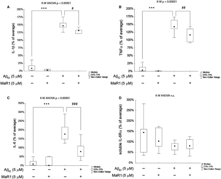
(A‐D) MaR1 reduced Aβ42‐induced secretion of pro‐inflammatory cytokines in d‐THP‐1 cells. Differentiated THP‐1 (d‐THP‐1) cells were incubated with vehicle, 5 µM Aβ42, 5 µmol/L MaR1 or 5 µM Aβ42 + 5 µM MaR1 for 24 h and the supernatants were analysed by ELISA. A total of 14 experiments were performed. MaR1 reduced the Aβ42‐induced increase in interleukin (IL)‐1β (A), tumour necrosis factor (TNF)‐α (B) and IL‐6 (C). The levels of IL‐6 receptor (R) α (D) were not affected by Aβ42 nor MaR1. Analysis of variance (ANOVA) was performed with the non‐parametric Kruskal–Wallis (K‐W) test, using the built‐in post hoc test for multiple comparisons to find significant differences between treatments. ***P < 0.005 vs. vehicle. # P < 0.05, ## P < 0.01, ### P < 0.005 vs. 5 μM Aβ42. Aβ = β‐amyloid; MaR1 = maresin 1
