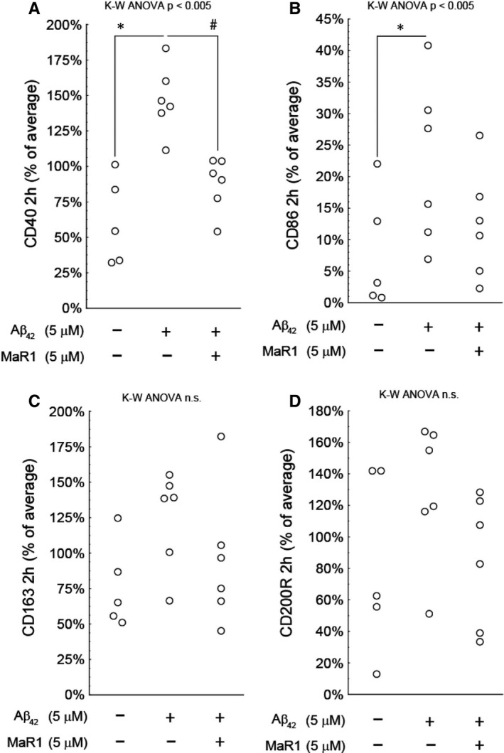
Figure 7.

(A‐D) MaR1 reduced pro‐inflammatory surface biomarkers. Differentiated THP‐1 (d‐THP‐1) cells were incubated for 2 h with 5 μM Aβ42 alone or together with 5 μM MaR1. Incubation with vehicle served as control. The percentage of cells expressing pro‐ and anti‐inflammatory surface biomarkers CD40, CD86, CD163 and CD200R was assessed by flow cytometry in a total of six individual experiments. Incubation with Aβ42 significantly increased the pro‐inflammatory biomarkers CD40 and CD86. MaR1 attenuated the increase in CD40 (P < 0.05), whereas the anti‐inflammatory markers CD163 or CD200R were not affected by Aβ42 nor by Aβ42 + MaR1. Analysis of variance (ANOVA) was performed with the non‐parametric Kruskal–Wallis (K‐W) test, using the built‐in post hoc test for multiple comparisons to find significant differences between treatments. ***P < 0.005 vs. vehicle. # P < 0.05 vs. 5 μM Aβ42. Aβ = β‐amyloid, MaR1 = maresin 1
