FIGURE 1.

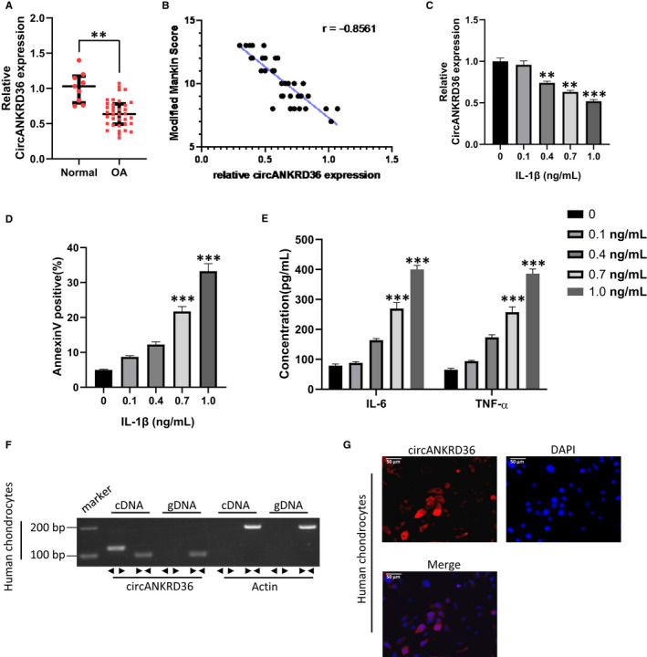
circANKRD36 is significantly decreased in osteoarthritis. A, Relative expression of circANKRD36 in OA (osteoarthritis) tissues (n = 36) and normal tissues (n = 9). **P < .01. B, Modified Mankin grading was performed to evaluate OA severity. (In the modified Mankin grading, abnormalities in structure (0‐6 points), cellularity (0‐3 points) and Safranin‐O staining (0‐4 points) were assessed up to a maximum score of 13 points.) The expression of circANKRD36 was negatively correlated with modified Mankin scores). C, Relative expression of circANKRD36 in chondrocytes treated with IL‐1β in a dose‐dependent manner (0, 0.1, 0.4, 0.7, 1.0 ng/mL). Data are presented as means ± SD (n = 3). **P < .01, ***P < .001. D, Human chondrocytes were treated with different doses of IL‐1β. Apoptotic cells were analysed by FACS with Annexin V staining. Annexin V–positive cell rates were statistically analysed. Data are presented as means ± SD (n = 3). **P < .01, ***P < .001. E, Human chondrocytes were treated with different doses of IL‐1β. Concentration of cytokines in culture medium was measured by ELISA Kit. Data are presented as means ± SD (n = 3). **P < .01, ***P < .001. F, The presence of circANKRD36 was validated in chondrocytes by RT‐PCR. Divergent primers amplified circANKRD36 from cDNA, but not from genomic DNA. GAPDH was used as a negative control. G, RNA FISH revealed the predominant localization of circANKRD36 in the cytoplasm. circANKRD36 probes were labelled with Cy‐3. Nuclei were stained with DAPI. Scale bar, 50 μm
