
Figure 2.
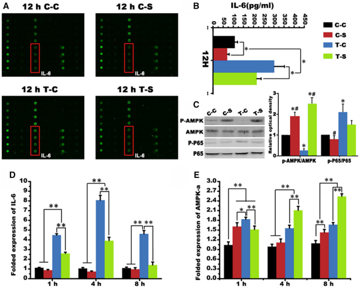
Protein secretion of PDLCs. A, Human cytokine antibody array screening of supernatant derived from co‐stimulated PDLCs. B, IL‐6 expression in supernatants derived from indicated treatment. C, The Western blot of AMPK and P65 and the folded relative optical dentistry of p‐AMPK/AMPK and p‐P65/ P65 (*, P < .05, ** P < .01 vs C‐C group; # P < .05, ##, P < .01 vs C‐S group; @, P < .05, @@, P < .01 vs T‐C group). D, Folded expression of IL‐6 indicated by Real‐time PCR. E, Folded expression of AMPK‐ɑ mRNA (*, P < .05, ** P < .01)