
Figure 6.
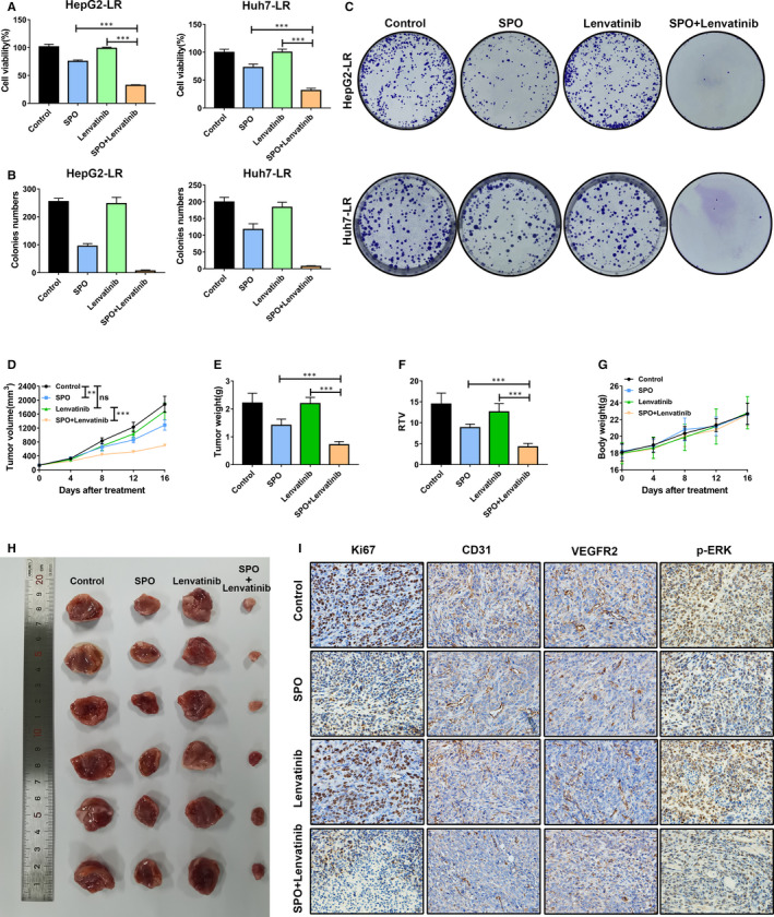
Sophoridine sensitized the anti‐tumour effect of lenvatinib against lenvatinib‐resistant HCC cell lines in vitro and in vivo. A, Lenvatinib‐resistant (LR) HepG2 or Huh7 cells were treated with 20 μmol/L Sophoridine combined with 5 μmol/L lenvatinib for 72 h. The combination treatment showed the most effective influence on the growth of HepG2‐LR and Huh7‐LR cells. B, The HepG2‐LR and Huh7‐LR cells were treated with 20 μmol/L Sophoridine combined with 5 μmol/L lenvatinib in colony formation assay. Colonies formation of HepG2‐LR and Huh7‐LR cells were inhibited after combination treatment. C, Representative images of the colony formation ability of HepG2‐LR and Huh7‐LR cells were shown after indicated treatments. D, BALB/c nude mice were subcutaneously burdened with HepG2‐LR cells. Mice were divided into different treatment groups after 7 days (n = 6). Mice were treated with Sophoridine (50 mg/kg, daily, intraperitoneally), lenvatinib (30 mg/kg, daily, intragastrically) or Sophoridine combined with lenvatinib. Tumour growth change curves were explored after indicated treatment. E, Tumour weight was detected after mice killing. F, The relative tumour volume (RTV) ratio was calculated according to the formula RTV = tumour volume day 16‐day 0/tumour volume day 0. G, Mouse bodyweight changes were observed during the treatments. H, The representative photograph of tumours was shown in different groups. I, Tumour slices of indicated groups were stained with Ki67, CD31, VEGFR2 and p‐ERK. The representative immunohistochemistry images of Ki67, CD31, VEGFR2 and p‐ERK from different groups were displayed. ns, P value > 0.05; **, P value < 0.01; ***, P value < 0.001