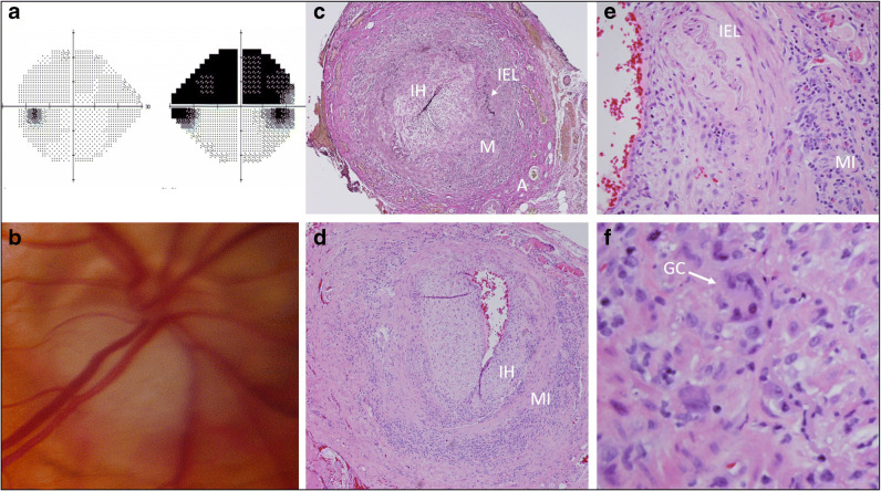
Fig. 3.
A case of giant cell arteritis. A 92-year-old woman complained of headache, jaw claudication, and vision loss. a Humphrey visual fields showed a superior altitudinal visual field loss in her right eye. b Funduscopy revealed a focal swelling inferiorly in the right optic disc that was also pale, i.e., pallid edema. Erythrocyte sedimentation rate (ESR) was 105 mm/h, C-reactive protein (CRP) was 3.6 dG/L. A temporal artery biopsy showed a mixed inflammatory infiltrate including giant cells within the arterial wall, consistent with GCA. c Elastin stain at low power demonstrate disruption of the internal elastic lamina (IEL) as well as a mixed infiltrate (MI) and intimal hyperplasia (IH). d Low power hematoxylin and eosin (H&E) demonstrates intimal hyperplasia (IH) and a mixed infiltrate (MI). e High power H&E demonstrates disruption of the IEL and a mixed infiltrate (MI). f High power H&E shows giant cells. (GC).

