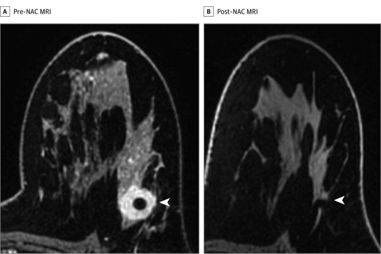
Figure 3. Magnetic Resonance Imaging (MRI) Before and After Neoadjuvant Chemotherapy (NAC) Demonstrating an Imaging Complete Response After NAC.
A, Axial fat-saturated T1-weighted postcontrast imaging of a biopsy-proven left breast ERBB2 (formerly HER2 or HER2/neu)–positive invasive ductal carcinoma (arrowhead). B, Axial fat-saturated T1-weighted postcontrast imaging of the same patient demonstrating an imaging complete response defined as no residual tumor enhancement in the treated tumor bed as indicated by the accurately positioned biopsy marker (arrowhead) and anatomic landmarks.

