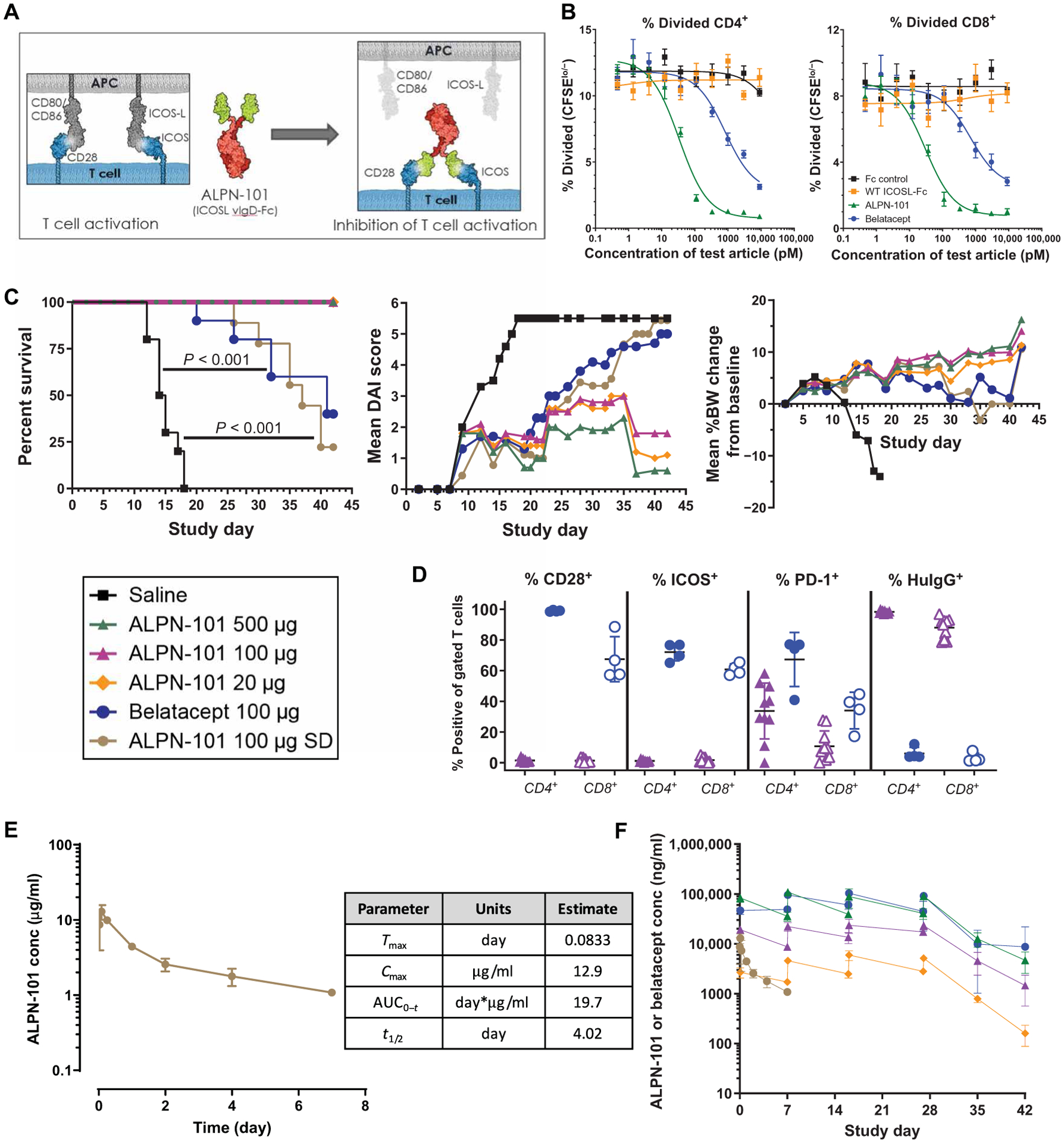
Fig. 3. ALPN-101 suppresses activated T cell expansion in the human PBMC-NSG GVHD model.

(A) Diagram of the structure of ALPN-101 and its mechanism of action. ALPN-101 was generated on the vIgD yeast display platform (21) and comprises two ICOSL “vIgD” domains (green) fused to a dimeric Fc tail (red) engineered to lack appreciable FcγR (CD16a, CD32, or CD64) or complement (C1q) binding while retaining FcRn binding. (B) Proliferation of CD4+ and CD8+ T cells was determined by quantifying the percentage of carboxyfluorescein diacetate succinimidyl ester (CFSE)–labeled cells remaining over time. As cells divide, CFSE signal decreases, and the percent CFSElo/− cells is used to assess the fraction of divided cells. Data are representative of at least six experiments with different donor pairs. (C) Survival, disease activity index (DAI) scores, and body weight (BW) of NSG mice x-ray irradiated (100 cGy) and administered 10 mg of human γ globulin subcutaneously on day −1 and then transplanted intravenously with 10 × 106 human PBMCs on day 0 and treated intraperitoneally with saline 3× per week (TIW) for 4 weeks (TIW × 4; days 0 to 28); 20, 100, or 500 μg of ALPN-101 TIW × 4; 100 μg of belatacept TIW × 4; or 100 μg of ALPN-101 single dose (SD) on day 0. On day 42, human CD45+ cells in blood were characterized by flow cytometry. For DAI analysis, the last observations or scores were carried forward for mice that were euthanized before the end of the study. For BW analysis, mice were weighed daily. See table S5 for statistical differences among groups. (D) Analysis of terminal blood collected from euthanized mice by flow cytometry for the 100 μg TIW × 4 ALPN-101 and belatacept treatment groups from the study described in (C). The % CD28+, ICOS+, PD-1+, or anti-human IgG-binding cells of gated CD4+ (filled) and CD8+ (open) from mice administered ALPN-101 (purple) or belatacept (blue). ALPN-101 blocks detection of CD28 and ICOS, and the anti-human IgG reagent binds to the Fc of cell-bound ALPN-101. Very few myeloid or B cells remained, so binding of belatacept to CD80/CD86 on APC was not detected. (E) Concentration (conc) of ALPN-101 in the serum of mice receiving a single dose of 100 μg of ALPN-101 in the GVHD model described in (C). Serum samples from three mice per group per time point after a single intraperitoneal injection of ALPN-101 were evaluated for drug concentrations by plate-based ELISA. (F) Concentrations of ALPN-101 and belatacept in the serum of mice receiving the indicated repeated doses (RD, TIW × 4) of the test articles in the GVHD model described in (C). Concentrations of ALPN-101 or belatacept were measured by ELISA in serum samples collected 2 hours post-dose on D0 (first dose), pre-dose, and 2 hours post-dose on D7, D16, and D27 (4th, 8th, and 12th doses) and D35 and D42 (8 and 15 days after the last dose). In (B), data are shown as mean ± SEM. In (C) (left), statistical significance was determined by Mantel-Cox log-rank test; in (C) (middle), by two-way repeated measures ANOVA for treatment effect; in (C) (right), by one-way ANOVA with Bonferroni correction. In (D), statistical significance was determined by unpaired t test.
