
Figure 1.
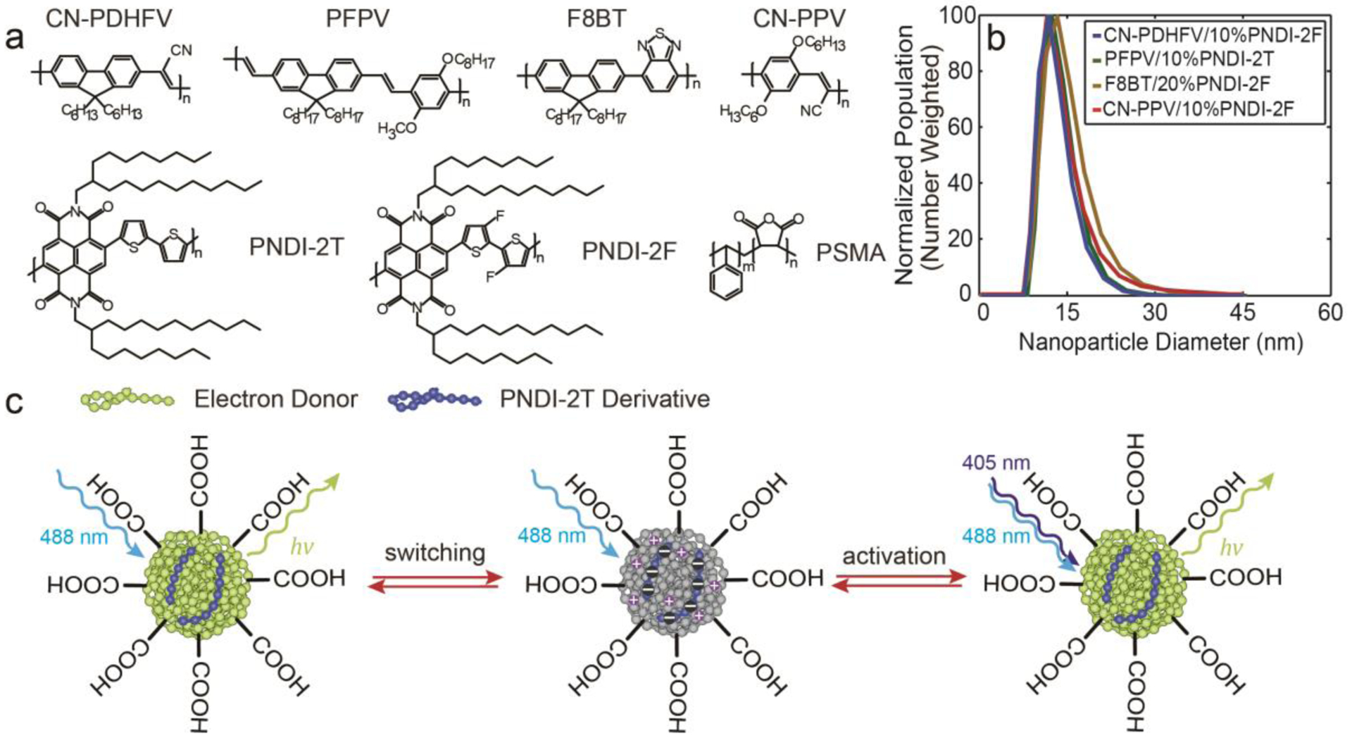
a) Chemical structures of CN-PDHFV, PFPV, F8BT, CN-PPV, PNDI-2T, PNDI-2F, and PSMA. b) Number-weighted particle size distributions of 10% PNDI-2F-doped CN-PDHFV Pdots, 10% PNDI-2T-doped PFPV Pdots, 20% PNDI-2F-doped F8BT Pdots, and 10% PNDI-2F-doped CN-PPV Pdots, determined using DLS. The corresponding particle sizes were 14.5±5.1 nm, 13.4±4.6 nm, 17.2±6.5 nm, 15.9±5.4 nm, respectively. c) Illustration of the photoswitching and photoactivation mechanisms of the Pdots.