Fig. 1.
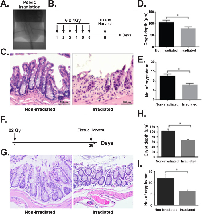
Radiation-induced damage to rectal tissue. a Portal camera image demonstrating pelvic irradiation (PIR) exposure field. b Schematic diagram demonstrating the timeline of fractionated pelvic irradiation. c H&E-stained representative cross section of the rectum. Non-irradiated mice showed a normal structure of crypts. However, mice exposed to fractionated pelvic irradiation suffer from crypt loss and epithelial damage. d, e Histogram demonstrating crypt depth (c) and number of crypt per mm (d) on rectal tissue. Exposure to fractionated pelvic irradiation significantly reduces crypt depth (d) (n 3, p < 0.0061, Mann-Whitney test) and number of crypt per mm (n 3, p value 0.0061, Mann-Whitney test) on rectal tissue (e). f Schematic diagram demonstrating the timeline of single fraction pelvic irradiation. g H&E-stained representative cross section of rectum. Non-irradiated mice showed a normal structure of crypts. However, mice exposed to pelvic radiation-induced epithelial damage with loss of crypt. h, i Histogram demonstrating mice exposed to pelvic irradiation decreases crypt depth (n 3, p < 0.0048, Mann-Whitney test) and crypt/mm (n 3, p < 0.0056, Mann-Whitney test) on rectal tissue

