Fig. 4.

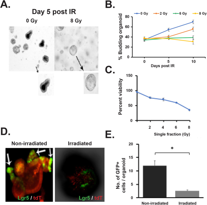
Effect of irradiation on rectal organoids. a, b Microscopic image (phase contrast) of rectal organoids along with histogram of % budding organoid demonstrating that irradiation impaired the organoid growth compared to un-irradiated control (2 Gy *p < 0.004, 6 Gy *p < 0.006). Microscopic image with × 10 (indicated with arrow) and × 40 magnification demonstrated loss of budding crypt in irradiated organoids. c Survival assay (ATP uptake assay) demonstrated significant reduction of organoid survival in a dose-dependent manner. d Confocal microscopic images of organoids developed from Lgr5-EGFP-CRE-ERT2; R26- ACTB-tdTomato-EGFP mice demonstrated loss of Lgr5+ve cells (Green) in budding crypt from irradiated organoids compared to un-irradiated control. tdTomato is constitutively expressed in these mice as membrane-bound protein therefore allows better visualization of cellular morphology. e Histogram demonstrating significant decrease in Lgr5 +ve cells in irradiated rectal organoids compared to un-irradiated control (p < 0.0004)
