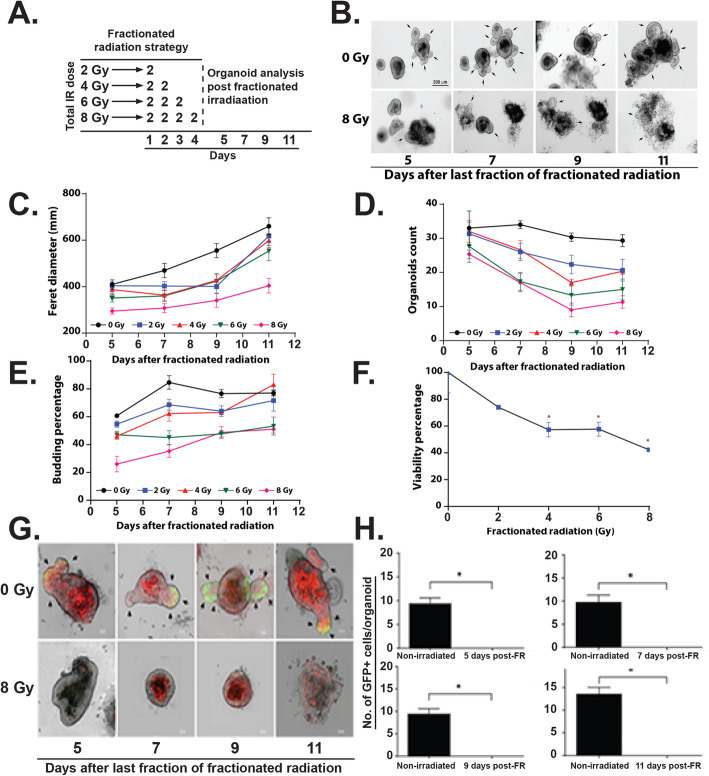
Fig. 5.
Effect of fractionated radiation on rectal organoids. a Schematic representation of the fractionated irradiation to organoid. b Microscopic image (phase contrast) of rectal organoids. c–e Significant decrease in physical parameters such as c Feret diameter (8 Gy vs 0 Gy (p < 0.0001) at day 8 post IR), d organoid count (8 Gy vs 0 Gy (p < 0.0028) at day 8 post IR), and e budding percentage (8 Gy vs 0 Gy (0.0094) at day 8 post IR) was observed in a dose-dependent manner. f Significant decrease in organoid viability was observed in a dose-dependent manner (8 Gy vs 0 Gy (0.0028) Mann-Whitney test). g Confocal microscopic images of organoids developed from Lgr5-EGFP-CRE-ERT2; R26-ACTB-tdTomato-EGFP mice demonstrated loss of Lgr5+ve cells (Green) in budding crypt from irradiated organoids compared to un-irradiated control. tdTomato is constitutively expressed in these mice as membrane-bound protein therefore allows better visualization of cellular morphology. h Histogram demonstrating significant decrease in Lgr5+ve cells in irradiated rectal organoids compared to un-irradiated control at different time point post irradiation (p < 0.0079, day 11 post IR)

