Fig. 3.
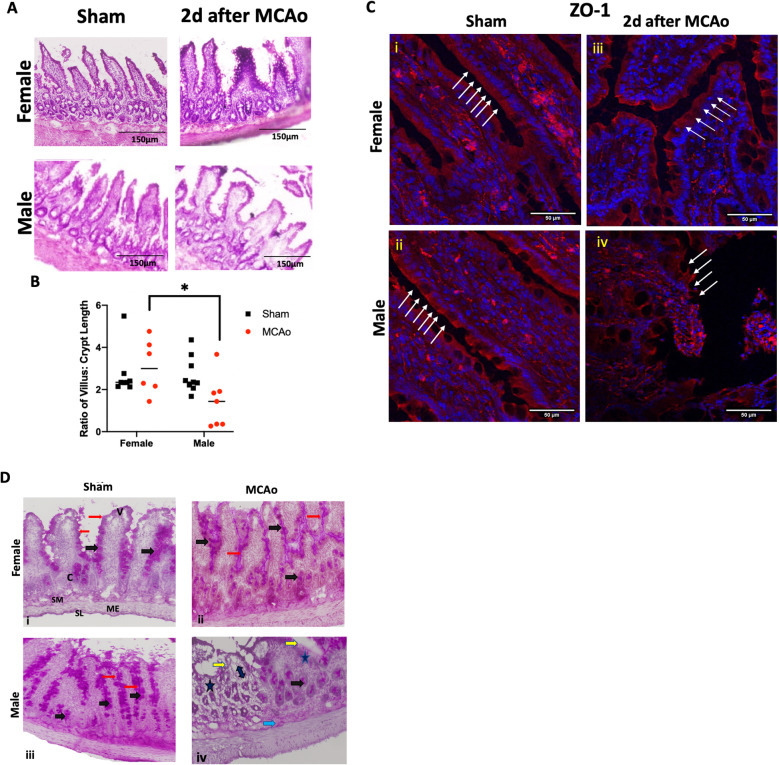
Histological analysis of the gut in sham and stroke animals. a H&E-stained sections of the ileum from sham and MCAo males and females. b Histogram depicting the mean (± SEM) ratio of villus:crypt height for each group. N = 7–9 per group, *p < 0.05. c Immunohistochemistry for the tight junction protein ZO-1 in thin sections of the ileum from male and female sham and MCAo groups. White arrows indicate the location of the epithelial barrier. Inter-epithelial expression of ZO-1 was virtually absent in male rats 2 days after MCAo compared to the other groups. Photomicrographs of the small intestine (ileum) showing periodic acid–Schiff (PAS) staining for mucin. In sham females (i), MCAo females (ii), and sham males (iii), a continuous and well-defined brush border of the villi (red arrows) is visible with strong pink color staining in the goblet cells (black arrows) of the villi and crypts. In MCAo males (iv), the brush border is interrupted (yellow arrows) with reduced mucin staining and reduction in goblet cells (asterisk) over the villi and crypts. Erosion of the gut mucosa is also seen in the core of the villi (double-headed arrow) as well as the submucosal layer (blue arrow). SM, submucosa; SL, serosa layer; ME, muscularis externa; V, villus; C, crypt

