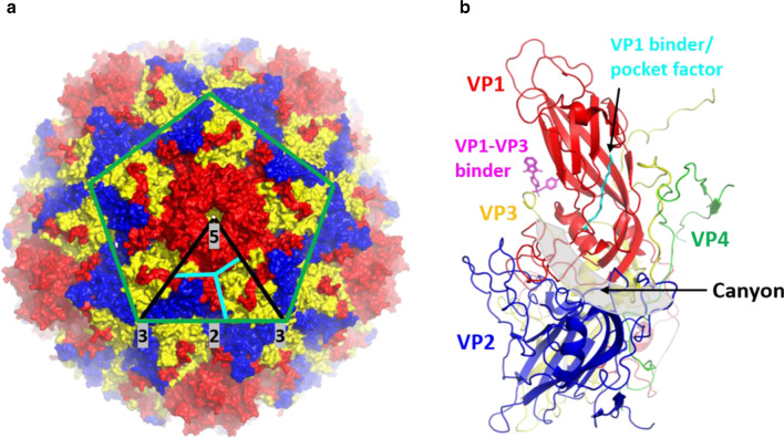
Fig. 1.
Enterovirus capsid organization and features. a Overall view of the enterovirus capsid comprising the VP1 (red), VP2 (blue) and VP3 (yellow). PDB ID: 4RQP [8]. The green lines indicate the boundaries of one pentamer. The black lines indicate the icosahedral symmetric subunit. The five, three, twofold symmetry axes are labeled and highlighted in grey. The cyan lines separate VP1 (red), VP2 (blue) and VP3 (yellow). Black arrows indicate the canyon region and the fivefold axis region formed by five VP1. b Canonical enterovirus protomer formed by VP1 (red), VP2 (blue), VP3 (yellow) and VP4 (green). PDB ID: 6GZV [9]. The canyon is highlighted in grey transparent and is indicated by a black arrow. Antiviral compounds that bind to the two binding pockets which are VP1 hydrophobic pocket and VP1–VP3 interprotomer pocket are shown in cyan and magenta, respectively

