Fig. 3.

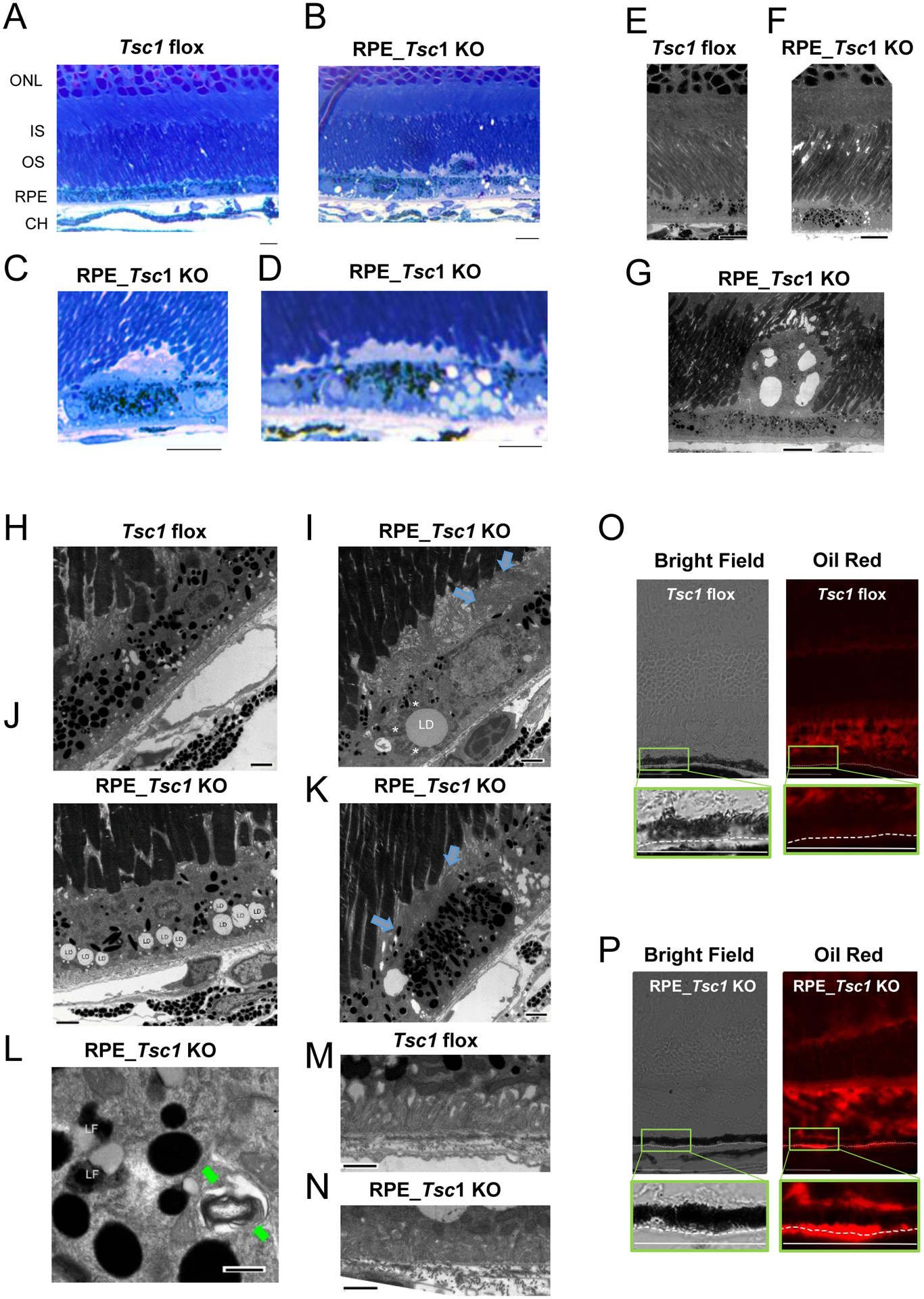
Lipid accumulation in the RPE of Tsc1 KO mice. (A-D) Toluidine blue-stained semi-thin sections of the posterior eyes from Tsc1 flox and RPE_Tsc1 KO at 4 ~ 5 months. The latter had cell infiltration (B) and accumulation of amorphous materials (C) on the apical face of the RPE, and accumulation of LD (D). (E to N) Transmission electron microscopy images of mice at 4 ~ 5 M. RPE-specific Tsc1 KO showed subretinal cell infiltration (G), membrane whorls on the apical faces of the RPE (blue arrows in I and K), LD with abnormal size (I) or abundance (J), preidroplet mitochondria (L, *), lipofuscin (LF) (L), undigested POS structure (L, green arrows), and mild disorganized basal infoldings (N). RPE hypo- (I) and hyper-pigmentation (K) were also detected. (O) and (P) Oil Red O staining of neural lipids on cryosections of posterior eyes prepared from RPE_Tsc1 KO animals and the Tsc1 flox counterparts. Scale bar: 10 μm (A-G), 2 μm (H-K), 1 μm (M and N), 400 nm (L), and 50 μm (O and P).
