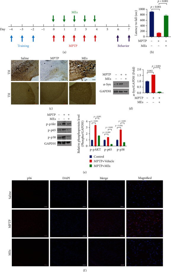
Figure 5.

MEs improve parkinsonism in MPTP-treated mice. (a) Experimental timeline for the construction of the MPTP-induced PD mouse model and the administration of MEs. C57BL/6 mice (male, 7-8-week-old) were injected MPTP intraperitoneally at 30 mg/kg/day for five days, and MEs (30 mg/kg/day) were injected intraperitoneally for 5 days since the administration of MPTP. On day 6, the rotarod test was performed. On day 7, mice were sacrificed and tissues were prepared for immunohistochemical (IHC) and western blotting. (b) Rotarod behavioral performance of MPTP-induced PD mice after ME treatment. Data are presented as the mean ± SD (n = 6). (c) Immunohistochemistry for tyrosine hydroxylase (TH) in the substantia nigra (scale bar = 100 μm) and striatum (scale bar = 1000 μm). (d) Expression of α-syn by western blot analysis. The blots were reprobed to detect GAPDH as the internal control. (e) Effect of MEs on the LPS-induced phosphorylation of Akt, p65, and p38 in the striatum. The protein in obtained tissue was analyzed and quantified by western blotting. (f) Effect of MEs on the nuclear translocation of NF-κB in substantia nigra (scale bar = 100 μm). C57BL/6 mice (male, 7-8-week-old) were injected MPTP intraperitoneally at 30 mg/kg/day for five days, and MEs (30 mg/kg) were injected intraperitoneally for 5 days since the administration of MPTP. On day 7, mice were sacrificed and substantia nigra tissues were collected, and immunofluorescence was performed.
