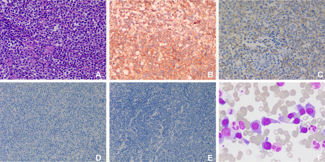
Figure 3.
Histopathological examination of masses in the breast and chest wall. (A) HE staining shows lymphocyte proliferation (original magnification, ×200); immunohistochemical staining of the proliferated lymphocytes show positive for CD138 (B, original magnification, ×200) and IgD (C, original magnification, ×200), but negative for CD56 (D, original magnification, ×100) and CD117 (E, original magnification, ×100); (F) bone marrow smears (original magnification, ×1000) show proplasmocytes and immature plasma cells, which differ in size and shape. The cytoplasm was dark blue with a few granules. The nucleus is biased and binuclear with reticular chromatin and vacuoles in some nuclei and plasma.

