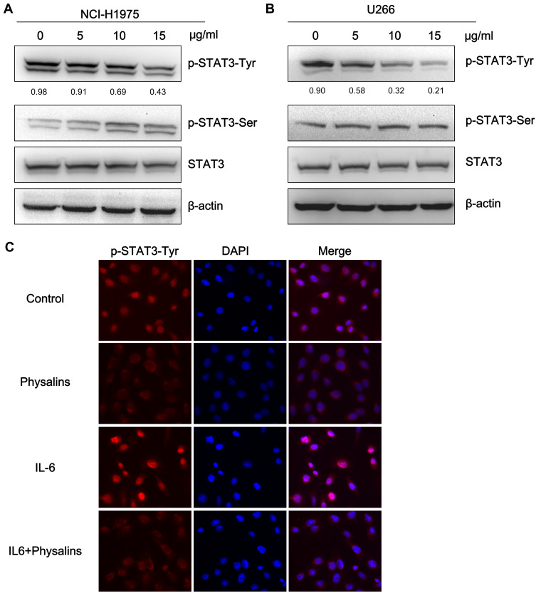
Figure 3.
Physalis alkekengi var. franchetii extracts suppressed constitutive STAT3 activity, and IL-6-induced STAT3 Tyr705 phosphorylation in NCI-H1975 and U266 cells. (A) NCI-H1975 cells were treated for 4 h with physalins (0, 5, 10, 15 μg/mL). The levels of p-STAT3-Tyr, p-STAT3-Ser, STAT3 and β-actin were detected by Western blot analysis. (B) U266 cells were treated for 4 h with physalins in a dose-dependent manner. The cell lysates were subjected to a Western blot analysis using antibodies specific for p-STAT3-Tyr, p-STAT3-Ser, STAT3 and β-actin. The semiquantification of the protein levels was performed with Image J software. The relative gray values of p-STAT3-Tyr are shown below. (C) Physalins suppressed p-STAT3 nuclear translocation. H1975 cells were treated with 15 μg/mL of physalins for 6 h with or without 25 ng/mL IL-6. Immunofluorescence analysis was performed with an anti-p-STAT3-Tyr primary antibody followed by an anti-rabbit IgG Fab2 Alexa Fluor 555 antibody. Coverslipped slides were covered with anti-fade reagents with DAPI. The merged images show the overlay of red Alexa Fluor 555 and blue DAPI fluorescence.

