Fig. 5.
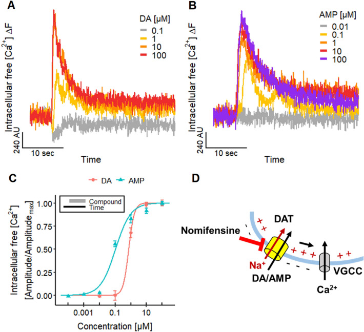
Altered neuronal signaling by DAT substrates. a, b Traces of Ca2+-imaging experiments illustrating the responses of the LUHMES neurons evoked by different concentrations of a dopamine (DA) and b amphetamine (AMP). c Concentration–response curves for DA and AMP with pEC50 values of 6.13 ± 0.12 and 6.98 ± 0.06, respectively. Note the treatment scheme (upper left corner), illustrating the experimental design. Detailed data on n numbers are found in table S4. d Schematic illustration of the underlying mechanisms of Ca2+-imaging signals evoked by DA and AMP. The transport of DA and AMP via the DAT into the cell leads to a net influx of one positive charge (Na+) that can active voltage-gated ion channels, like CaV ion channels. The DAT can be blocked by nomifensine

