Figure 16.

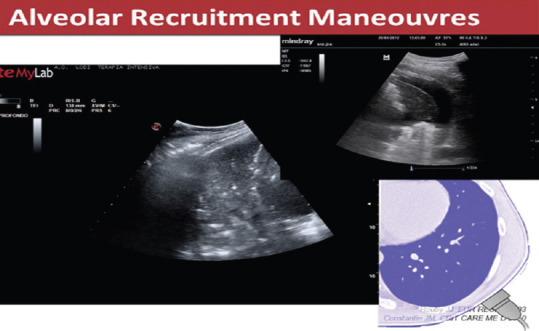
Lung consolidation with air bronchogram (seen as white spots on the left image) and a collapsed area surrounded by a pleural effusion (right image)

Lung consolidation with air bronchogram (seen as white spots on the left image) and a collapsed area surrounded by a pleural effusion (right image)