
Figure 2.
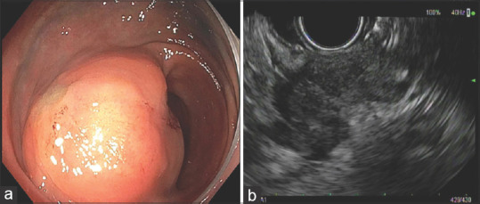
Recurrent rectal adenocarcinoma. (a) Endoscopic findings: Moderate sized, firm, subepithelial mass with smooth mucosa in the rectosigmoid region; (b) EUS findings: Hypoechoic mass within the sigmoid/rectal wall, with intact mucosa. The mass was involving the muscularis propria, submucosa. The mass had an infiltrating appearance and appeared to be invading into perirectal fat