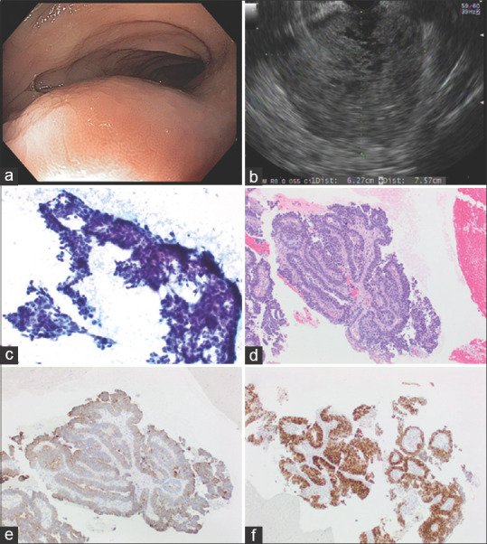
Figure 3.

Adenocarcinoma arising from endometriosis (a) Endoscopic findings: Smooth subepithelial compression without any intraluminal disease process. (b) EUS Findings: A large, 7.57 cm by 6.27 cm perirectosigmoid mass was seen that appeared to be contiguous with muscularis propria of the rectum in some views. The mass was hypoechoic but with cystic (anechoic) areas; (c) EUS-FNA sample: Papanicolaou stained direct smear; (d) H and E stained biopsy; (e) immunoperoxidase stain for cytokeratin 7 is positive in the tumor cells; (f) immunoperoxidase stain for estrogen receptor is positive in the tumor cells
