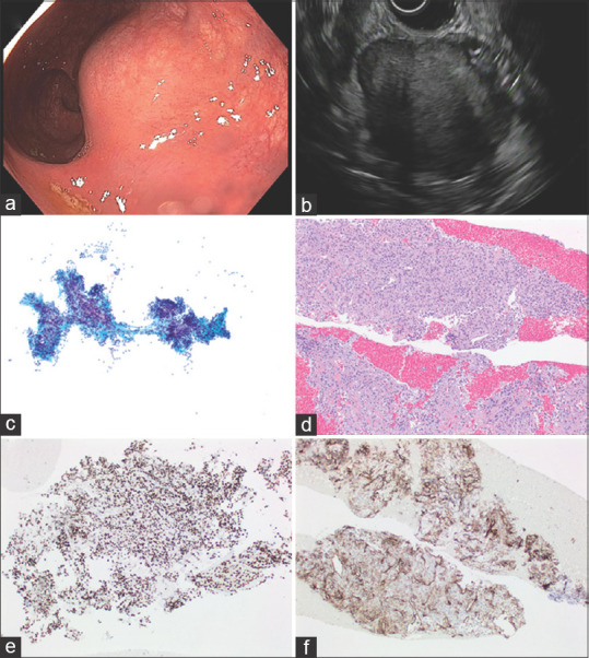
Figure 4.

Solitary fibrous tumor (a). Endoscopic findings: A large sub-epithelial mass found in the distal rectum measuring 5 cm in length (b) EUS: A hypoechoic, hyperechoic, and heterogeneous mass were found in the right-lateral perirectal space. The endosonographic borders were well-defined. The mass measured 50 mm (in maximum width) by 41 mm (in maximum thickness). The mass appeared to be contiguous with the muscularis propria suggesting that it is arising from the MP or invading it. (c) Papanicolaou stained FNA direct smear; (d) H and E stained biopsy; (e) immunoperoxidase stain for STAT6 is positive in the tumor cells; (f) immunoperoxidase stain for CD34 is positive in the tumor cells
