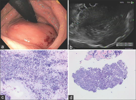
Figure 5.

Lymphoma (a) endoscopic findings: A subepithelial (with smooth mucosal surface) partially obstructing large mass was found in the rectum. (b) EUS: A hypoechoic and heterogeneous mass was found in the perirectal space. The mass was visualized endosonographically with the probe positioned at 0.5 cm (from the anal verge). The endosonographic borders were irregular; (c) Romanowsky stained FNA direct smear; (d) H and E stained cell block section of the FNA. Flow cytometric analysis demonstrated aberrant monoclonal B-cell population consistent with lymphoma
