Figure 1.

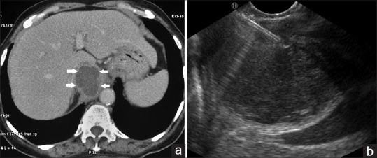
(a) Computed tomography abdomen: Amoebic liver abscess in caudate lobe (arrow). (b) EUS-guided drainage of liver abscess. The abscess being punctured with 19G needle

(a) Computed tomography abdomen: Amoebic liver abscess in caudate lobe (arrow). (b) EUS-guided drainage of liver abscess. The abscess being punctured with 19G needle