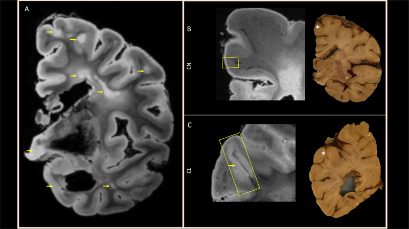
Fig. 1.
Preparation of the tissue for magnetic resonance microscopy (MRM): (A) Representative slice from T2* -weighted ex-vivo whole-hemisphere MRI depicts cortical, subcortical, and deep WM lesions (arrows). From images like these, small regions highlighted by the yellow boxes on zoomed-in ex-vivo MRI (left side of panels B and C) and localized with asterisks on photographs of the tissue slab surfaces (right side of panels B and C), encompassing (B) normal appearing cortex and WM (CN block) and (C) a leukocortical lesion (CL block), were excised for magnetic resonance microscopy (MRM). Note that the MRI image shown in (C) is from deep inside the 1-cm thick slab, but the cortical lesion is not visible on the slab surface.

