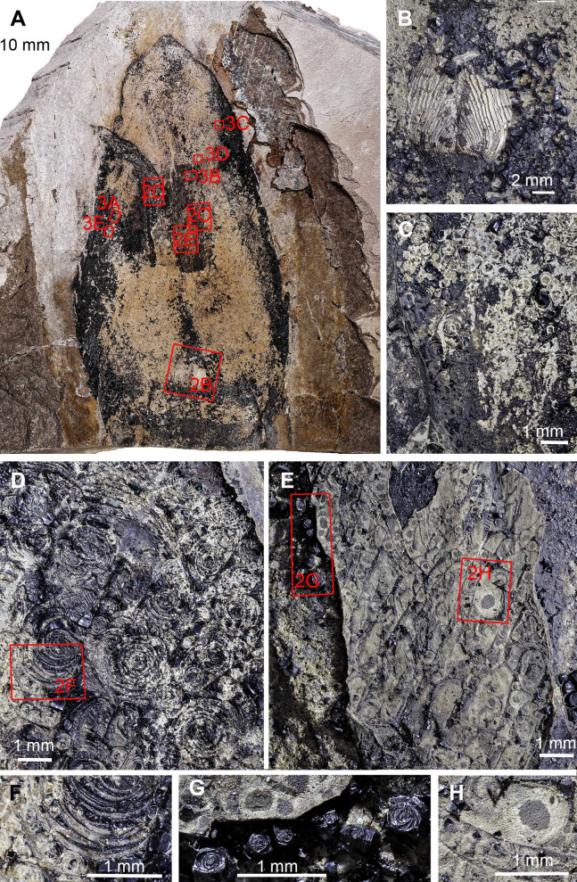
Figure 2. Trachyteuthis hastiformis, Kimmeridgian, Painten (Germany). SMNS 70496, leg. M. Kapitzke.
The details of A shown in B to H and Fig. 3 are marked with red rectangles. (A) Overview. (B) Detail of Lamellaptychus probably in the digestive tract of the animal. (C) Detail showing many light gray Type I-circles with dark gray centers. (D) Concentric Type II-circles rich in organic matter preserved like gagate. (E) Light Type I-circles like in C; note the linear arrangement. (F) Detail of (D) showing Type I- and II-circles of varying sizes (the largest is Type II). (G) Organic rich Type III-circles composed of mainly former ink (from E), with some Type I-circles at the top. (H) detail of the light gray Type I-circles in (E); apparently, the lighter outer ring may also have concentric structures, which are just not as distinct as when there was more ink between each subsequent ring. Photo credit: Dieter Buhl.

