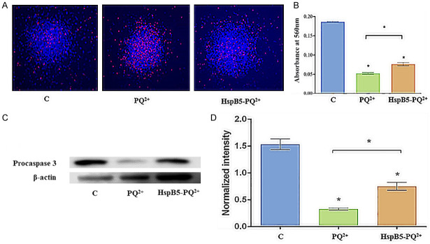
Figure 3.
Anti-apoptotic activity of HspB5 against paraquat-induced oxidative stress. A. mNSPCs were exposed to 3 mM paraquat for 4 h, stained with Hoechst (10 µg/mL) - PI (50 µg/mL) and observed under the fluorescence microscope. Here, [C] represents the control mNSPCs without any paraquat stress, [PQ2+] represents the mNSPCs treated with paraquat, and [HspB5-PQ2+] represents the mNSPCs pre-treated with HspB5 (100 µg/mL). Scale bar = 100 µm; B. Quantification of cell viability using the MTT assay; * represents statistical significance between the different treatment groups at p<0.001. C. Immunoblotting of apoptotic procaspase-3 in mNSPCs after paraquat stress. D. Western blot showing the expression of procaspase-3 expression in control, PQ2+ treated, and HspB5-PQ2+ treated. The densitometric measurements of all the detected proteins were performed by Scion NIH Image Analysis Software (Image J) (version 3.5). Each measurement represents the mean ± SD of the intensity of the bands normalized to their respective control (β-actin). A significant difference (p<0.001) was found between the different groups by paired 2-tailed Student’s t-test.

