Fig. 8.
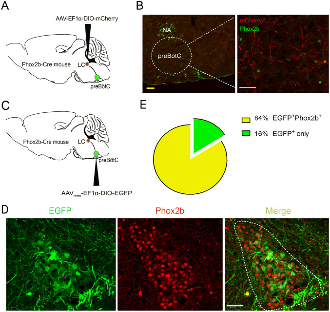
Projection of Phox2bLC neurons to the preBötC. A Schematic of anterograde virus injection into the LC of Phox2b-Cre mice. B Photomicrographs showing that mCherry-labeled axons of Phox2bLC neurons project to the preBötC region. Left: dashed line, contour of the preBötC; right: enlarged image showing Phox2b-labeled NA and RTN neurons (green). Scale bars, 50 μm. C Schematic of retrograde virus injection into the preBötC in Phox2b-Cre mice. D Confocal images showing the cell bodies of EGFP-expressing neurons in the LC, most of which are Phox2b+. E Cell counts showing that > 84% of EGFP neurons (n = 9 mice) projecting to the preBötC are Phox2b+ (scale bars, 50 μm).

