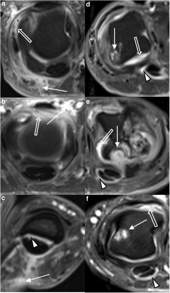
Fig. 3.
Detailed view of representative MRI findings in PsA and RA. Transversal T1w fat-saturated contrast-enhanced sequences of selected MCP joints of three patients each with PsA (a–c) and RA (d–f). a 34-year-old male. Severe periarticular inflammation (white arrow) with additional mild synovitis (open arrow). b 42-year-old female. Severe dorsal periarticular inflammation (white arrow) with synovitis (open arrow). c 44-year-old female. Severe periarticular inflammation (white arrow) with corresponding mild flexor tenosynovitis (arrowhead). d 39-year-old male. Widespread synovitis (white arrow) with moderate flexor tenosynovitis (arrowhead) and large bone erosion (white arrow). e 41-year-old male. Multiple large bone erosions (white arrow) and severe synovitis (open arrow) with mild flexor tenosynovitis (arrowhead). f 56-year-old female. Bone erosion (white arrow) with moderate flexor tenosynovitis (arrowhead) and moderate synovitis (open arrow). Note the absence of periarticular inflammation in d–f despite significant inflammatory joint changes at the joint level

