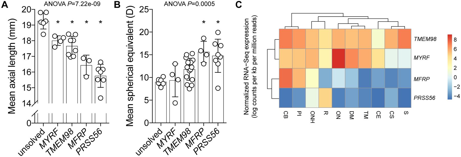
Figure 3: Clinical and transcriptional phenotypes of dominant and recessive forms of nanophthalmos.

Mean ocular axial length (A) and spherical equivalent (B) of affected individuals stratified by genetic diagnosis. For both phenotypes, group means were significantly different by one-way ANOVA (P<0.0005), with asterisks (*) indicating a significant difference from the unsolved cohort (Tukey multiple comparison testing, P<0.03). (C) Relative mean expression dendrogram (normalised log counts per kb per million mapped reads) of genes in dissected human adult cadaveric eye tissue. S, sclera; CS, corneal stroma; CE, corneal epithelium; TM, trabecular meshwork; DM, Descemet’s membrane; ON, optic nerve; ONH, optic nerve head; PI, peripheral iris; CB, ciliary body.
