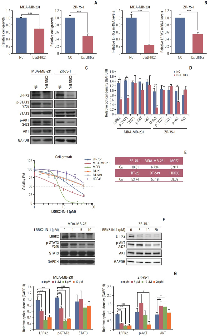
Fig. 6.
Inhibition of LRRK2 in LRRK2-overexpressing breast cancer cell lines reduces cell viability. (A) Suppression of LRRK2 expression leads to reduced cell growth in MDA-MB 231 and ZR-75-1 cells. (B, C) The efficiency of LRRK2 knockdown is evaluated by quantitative reverse transcription–polymerase chain reaction and western blotting. (D) Data quantification of panel (C). (E) Breast cancer cell lines overexpressing LRRK2 respond to LRRK2-IN-1 dose-dependently. (F) Immunoblot of LRRK2-IN-1-treated MDA-MB-231 and ZR-75-1 cells. The blots of individual cell lines crop from different part of the same gel, respectively. The ZR-75-1 cell lines data of LRRK2-IN-1 were captured by an ImageQuant LAS 4000 biomolecular imager. (G) Data quantification of panel (F). GAPDH, glyceraldehyde 3-phosphate dehydrogenase; NC, negative control siRNA. Values are presented as mean±standard deviation. *p < 0.05, **p < 0.01, ***p < 0.001.

