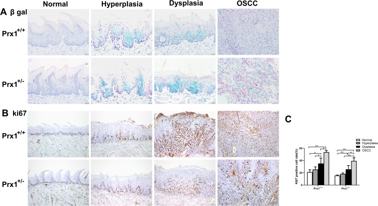
Figure 2.
The levels of cellular senescence in tongue tissues from Prx1+/+ and Prx1+/- mice. (A) Representative β-gal staining in tongue tissues from Prx1+/+ and Prx1+/- mice in each group. (B and C) In each group, Ki67 was measured in the tongue tissues by IHC and the percentage of Ki67-positive cells was analyzed (magnification: 200×, data represent mean values ± SD, *p <0.05, **p<0.01).

