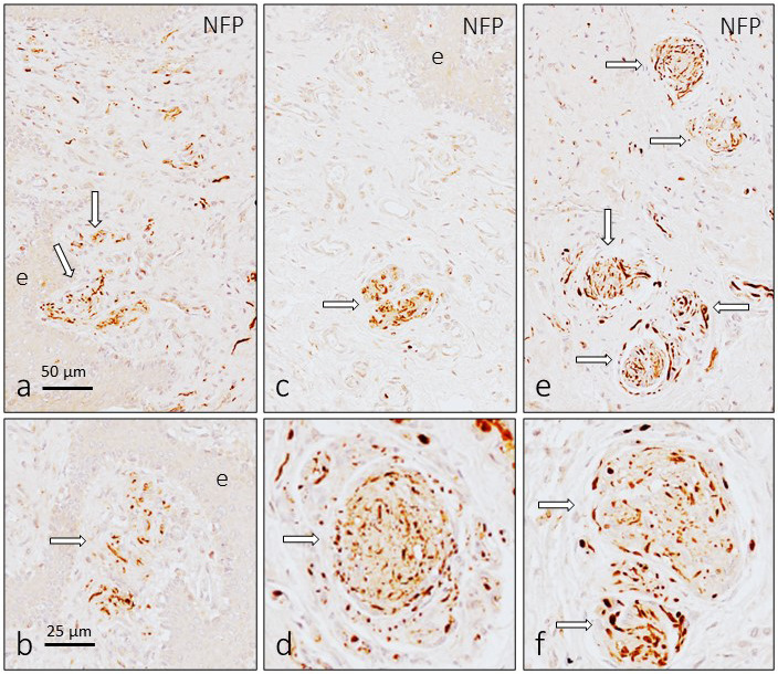
Figure 3.

Immunohistochemical detection of neurofilament protein and neuron‐specific enolase in the sensory corpuscles of the human glans clitoris. The arrangement of the axons in the dermal papillae was similar to that of cutaneous Meissner corpuscles (a and b), whereas in those placed in deep subepithelial tissues and in the central part of the organ (c–f), the axon was very tightly coiled and look like a “wool ball” or “yarn ball” with remarkable differences in the diameter of the axonal profiles
