Figure 8.

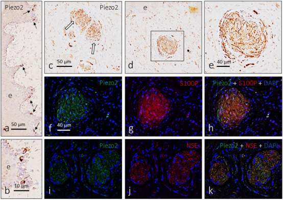
Immunodetection of PIEZO2 in the glans clitoris. PIEZO2 immunoreactivity is detected in cells of the epithelium basal layer (a and b) that were identified as Merkel cells (arrows in a). In the genital endbulbs, PIEZO2 shows a typical “wool ball” or “yarn ball” axonal pattern of distribution (c–e; arrows in c). Immunofluorescence confirmed that PIEZO2 is localized in the axon (i–k) and is absent from the Schwann‐related cells (f–h)
