Figure 1.

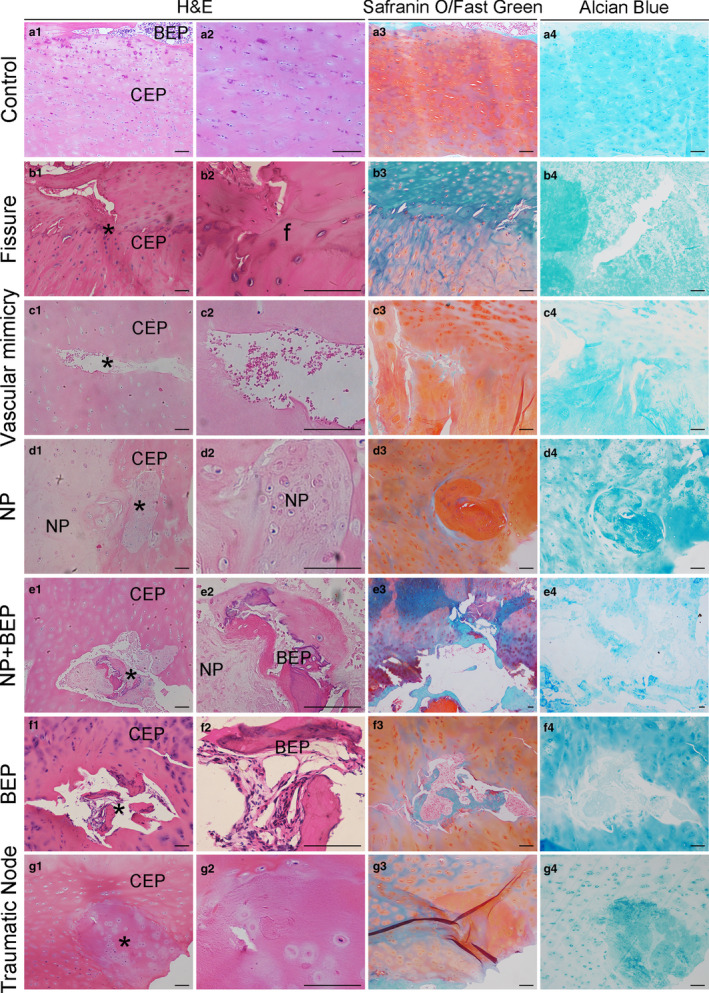
Histology of cartilage endplates (CEPs). (a1–a4) Control (normal) CEPs stained with H&E, Safranin O/Fast green, and Alcian Blue staining. Some bony endplate (BEP) is seen at the top of some images. (b1–b4) CEPs with fissures. The cartilage calcification ‘tidemark’ lies centrally in some images. (c1–c4) CEPs with ‘vascular mimicry’ suggesting blood vessel ingrowth after damage. (d1–d4) Nucleus pulposus (NP) herniation into the CEPs. (e1–e4) NP herniation and incorporation of bone tissue into the CEPs. (f1–f4) Incorporation of bone tissue into the CEPs. (g1–g4) The traumatic nodes (damaged/repaired CEPs) within CEPs. BEP, bony endplate; CEP, cartilaginous endplate; NP, nucleus pulposus; f, fissure; *The position of typical structural features. Scale bar: 100 μm
